

Association of localized intravascular coagulopathy with venous malformations
- PMID: 18645138
- PMCID: PMC5572565
- DOI: 10.1001/archderm.144.7.873
Association of localized intravascular coagulopathy with venous malformations
Abstract
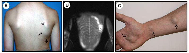
Objective: To determine which venous malformations (VMs) are at risk for coagulopathy. Venous malformations are slow-flow vascular malformations present at birth, and localized intravascular coagulopathy (LIC) causes pain and thrombosis within a lesion and severe bleeding during surgical procedures.
Design: Prospective convenience sample accrued from 2 multidisciplinary sites in Brussels, Belgium, and Caen, France.
Participants: The study population comprised 140 patients with clinical data and coagulation parameters. Magnetic resonance imaging was performed for 110 patients.
Main outcome measure: Measurement of D-dimer levels.
Results: Of the 140 participants, 59 (42%) showed high D-dimer levels, 36 (61%) of whom had levels higher than 1.0 microg/mL. Six of the participants had low fibrinogen levels. In univariate analysis, large surface, presence of palpable phleboliths, and truncal localization were associated with high D-dimer levels. In the multivariate analysis, only large surface area and presence of phleboliths remained independently associated with high D-dimer levels. Severe LIC, characterized by concomitant low fibrinogen level, was associated with extensive venous malformations of the extremities.
Conclusions: Localized intravascular coagulopathy is statistically significantly associated with large and/or deep venous malformations that affect any location, which can have a palpable phlebolith. These patients are at risk of local pain due to thrombosis. Lesions with elevated D-dimer levels associated with low fibrinogen levels (severe LIC) commonly affect an extremity and have a high risk of hemorrhage. Low-molecular-weight heparin can be used both to treat the pain caused by LIC and to prevent decompensation of severe LIC to disseminated intravascular coagulopathy.
Figures
Comment in
-
The hidden face of venous malformations: a multidisciplinary therapeutic approach.Arch Dermatol. 2008 Jul;144(7):922-6. doi: 10.1001/archderm.144.7.922. Arch Dermatol. 2008. PMID: 18645144 No abstract available.
-
Oral contraceptive and D-dimer level.Arch Dermatol. 2009 Feb;145(2):210; author reply 210-1. doi: 10.1001/archdermatol.2008.565. Arch Dermatol. 2009. PMID: 19221279 No abstract available.
References
-
- Mulliken JB, Glowaki J. Hemangiomas and vascular malformations in infants and children : a classification based on endothelial characteristics. Plast Reconstr Surg. 1982;69:412–420. - PubMed
-
- Boon LM, Mulliken JB, Enjolras O, Vikkula M. Glomuvenous malformations (glomangioma) and venous malformation Distinct clinicopathologic and genetic entities. Arch Dermatol. 2004;140:971–976. - PubMed
-
- Vikkula M, Boon LM, Mulliken JB. Molecular basis of vascular anomalies. Trends Cardiovasc Med. 1998;8:281–292. - PubMed
-
- Brouillard P, Vikkula M. Vascular malformations : localized defects in vascular morphogenesis. Clin Genet. 2003 May;63(5):340–51. - PubMed
-
- Enjolras O, Mulliken JB. The current management of vascular birthmarks. Pediatr Dermatol. 1993;10:311–333. - PubMed
Publication types
MeSH terms
Substances
Grants and funding
LinkOut - more resources
Full Text Sources
Other Literature Sources
Medical
