

doi: 10.1107/S0907444908017393.
Epub 2008 Jul 17.
Representation of viruses in the remediated PDB archive
Affiliations
- PMID: 18645236
- PMCID: PMC2677383
- DOI: 10.1107/S0907444908017393
Item in Clipboard
Representation of viruses in the remediated PDB archive
Acta Crystallogr D Biol Crystallogr.
2008 Aug.
Abstract
A new scheme has been devised to represent viruses and other biological assemblies with regular noncrystallographic symmetry in the Protein Data Bank (PDB). The scheme describes existing and anticipated PDB entries of this type using generalized descriptions of deposited and experimental coordinate frames, symmetry and frame transformations. A simplified notation has been adopted to express the symmetry generation of assemblies from deposited coordinates and matrix operations describing the required point, helical or crystallographic symmetry. Complete correct information for building full assemblies, subassemblies and crystal asymmetric units of all virus entries is now available in the remediated PDB archive.
Figures
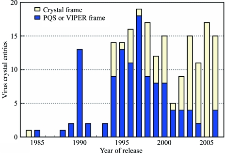
Deposition frame of remediated icosahedral virus crystal structure entries. The number of entries is plotted by year of release and coordinate frame type. Entries with coordinates provided in the standard frame of the crystal lattice are represented by light yellow bars. Entries presented in an icosahedral frame and requiring one or more non-identity transformations to place virus particles into the crystal lattice are represented by dark blue bars.

Examples of remediated PDB entries with regular noncrystallographic symmetry. (a) 1f2n , yellow mottle virus with icosahedral symmetry (Qu et al., 2000 ▶). (b) 4rhv , rhinovirus with icosahedral symmetry (Arnold & Rossmann, 1988 ▶). In (a) and (b), the icosahedral asymmetric unit is shown in ribbon representation. (c) 2bk1 , viral toxin pneumolysin with C38 circular symmetry (Tilley et al., 2005 ▶). (d) 1f2n , clathrin cage with D6 symmetry (Fotin et al., 2006 ▶). (e) 1ei7 , tobacco mosaic virus coat protein four-layer aggregate with D17 symmetry (Bhyravbhatla et al., 1998 ▶). (f) 1cgm , cucumber green mottle mosaic virus (CGMMV) with helical symmetry (Wang & Stubbs, 1994 ▶). Nucleic acid positions are shown in green and red. (g) 1ifd , filamentous phage with helical symmetry and fivefold circular symmetry (Marvin, 1990 ▶). Each color represents a strand winding about the helical axis. (h) 1m4x , P. bursaria chorella virus type 1 (PBCV-1) algal virus shell (Nandhagopal et al., 2002 ▶). Colors highlight pentasymmetron units (cyan) and trisymmetron units (red, yellow or blue–green–magenta). (i) Adjacent PBCV-1 pentasymmetron and trisymmetron. The position of the deposited coordinates for the protein trimer is shown in yellow. The subassembly corresponding to the icosahedral point asymmetric unit (one fifth of the pentasymmetron plus one third of the trisymmetron) is outlined in gray.
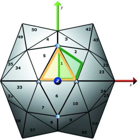
Icosahedral standard frame, shown with respect to orthogonal coordinate axes. Fivefolds and threefolds nearest to the the z axis are identified with symbols. Numbers show the order of symmetry operations for positions visible in this view. Yellow and green lines delimit the two alternate restricted placement boundaries for the first point asymmetric unit position.
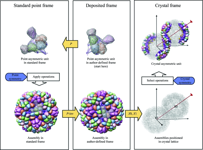
Assembly generation with regular point-symmetry example: 1al0 , crystal structure of ϕX174 procapsid (Dokland et al., 1997 ▶). The pathway to generate assemblies in standard point, author-defined and crystal frames is shown. Frame transformations are represented by yellow arrows connecting the deposited frame, standard icosahedral point frame and crystal frame. See §3.3 for details.
References
Publication types
MeSH terms
Grants and funding
LinkOut - more resources
Full Text Sources
