

doi: 10.3904/kjim.2008.23.2.106.
Dynamic left ventricular outflow tract obstruction without basal septal hypertrophy, caused by catecholamine therapy and volume depletion
Affiliations
- PMID: 18646515
- PMCID: PMC2686978
- DOI: 10.3904/kjim.2008.23.2.106
Item in Clipboard
Dynamic left ventricular outflow tract obstruction without basal septal hypertrophy, caused by catecholamine therapy and volume depletion
Korean J Intern Med.
2008 Jun.
Abstract
Hypertrophic cardiomyopathy (HCM) with hypertrophy of the basal septum is the most common etiology of left ventricular outflow tract (LVOT) obstruction. In this article, we report the case of a patient with a structurally normal heart who developed hemodynamic deterioration due to severe LVOT obstruction following treatment with catecholamines. Hypovolemia accompanied with a hyperdynamic condition, resulting from catecholamine treatment, may cause dynamic LVOT obstruction due to the systolic anterior motion of the mitral valve leaflet. The solution for this is early recognition and correction of aggravating factors such as, withdrawal of catecholamine therapy and volume replacement.
Figures
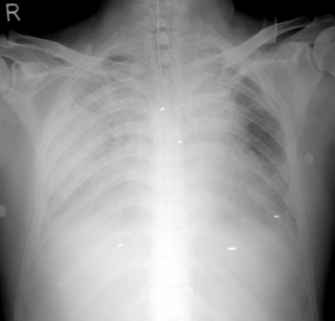
Chest X-ray showed diffuse reticulonodular opacities in both lungs and pulmonary edema.
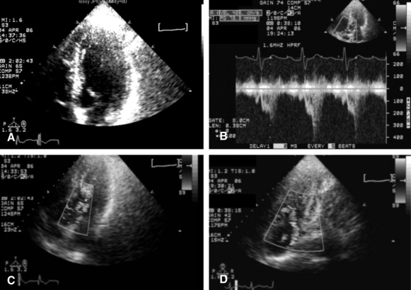
Transthoracic echocardiogram. (A) Systolic anterior motion of the mitral leaflets with left ventricular outflow tract obstruction was shown. (B) The peak pressure gradient measured with continuous Doppler in LVOT was 73.3 mm Hg. (C) Transthoracic echocardiography with color Doppler imaging showed flow acceleration in LVOT. (D) Moderate amount of mitral regurgitation was revealed on color Doppler examination.
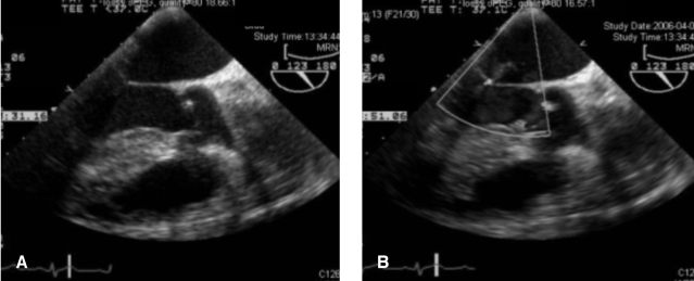
Transesophageal echocardiography was done to evaluate heart function after the treatment, i.e., volume replacement and stop of catecholamine infusion and to rule out acute derangement from infective endocarditis. (A) There was no more systolic anterior motion of the mitral valve. (B) The amount of mitral regurgitation reduced to trivial degree.
References
-
- Maron BJ, Bonow RO, Cannon RO, 3rd, Leon MB, Epstein SE. Hypertrophic cardiomyopathy: interrelations of clinical manifestations, pathophysiology, and therapy (2) N Engl J Med. 1987;316:844–852. - PubMed
-
- Heo J, Jeong JW, Park YK, Park OK. Relationship between systolic anterior motion of the mitral valve and the left ventricular outflow pressure gradient in patients with hypertrophic obstructive cardiomyopathy. Korean Circ J. 1990;20:351–357.
-
- Kim KS, Jung HY, Jo JH, Kim MS, Song JS, Bae JJ. A clinical study of hypertrophic obstructive cariomyopathy: analysis by echocardiography and Doppler echocardiography. Korean Circ J. 1988;18:647–656.
-
- Gallet B, Quintard H, Fruchaud J, Hiltgen M. Dynamic left ventricular obstruction associated with a pheochromocytoma reversible after tumor ablation. Arch Mal Coeur Vaiss. 2001;94:1195–1198. - PubMed
-
- Auer J, Berent R, Weber T, Lamm G, Eber B. Catecholamine therapy inducing dynamic left ventricular outflow tract obstruction. Int J Cardiol. 2005;101:325–328. - PubMed
MeSH terms
Substances
LinkOut - more resources
Full Text Sources