Review
. 2008:10:391-411.
doi: 10.1146/annurev.bioeng.9.060906.151929.

Chemical exchange saturation transfer contrast agents for magnetic resonance imaging

Affiliations
Review

Chemical exchange saturation transfer contrast agents for magnetic resonance imaging

A Dean Sherry et al. Annu Rev Biomed Eng. 2008.

Abstract

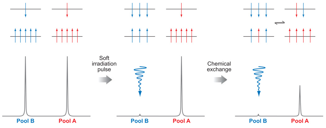
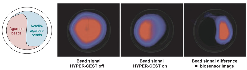
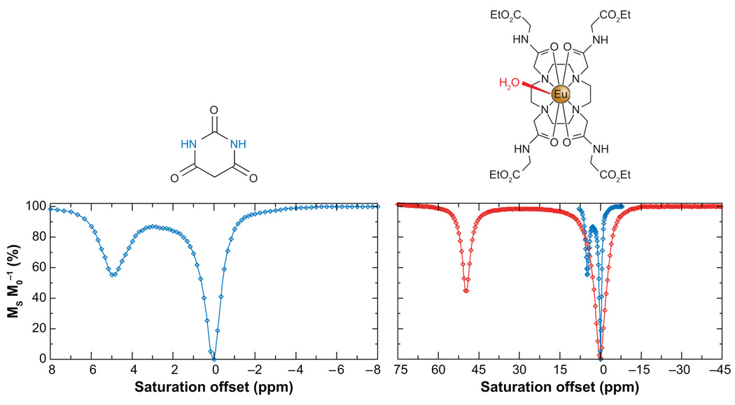
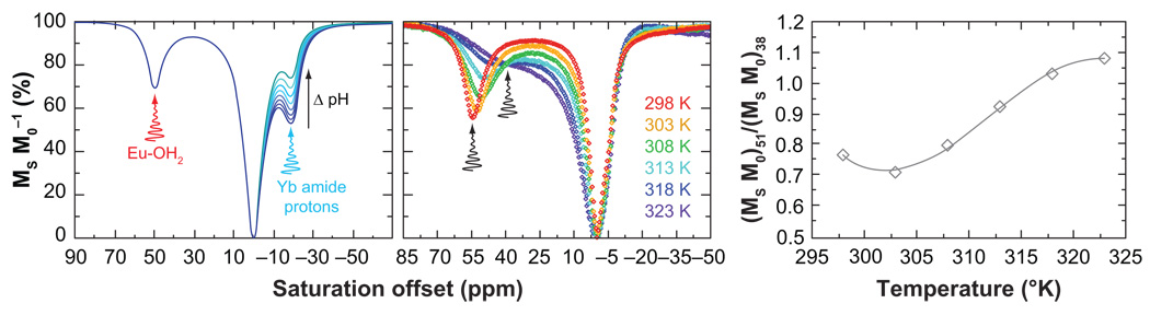
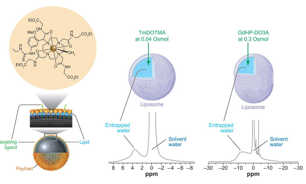
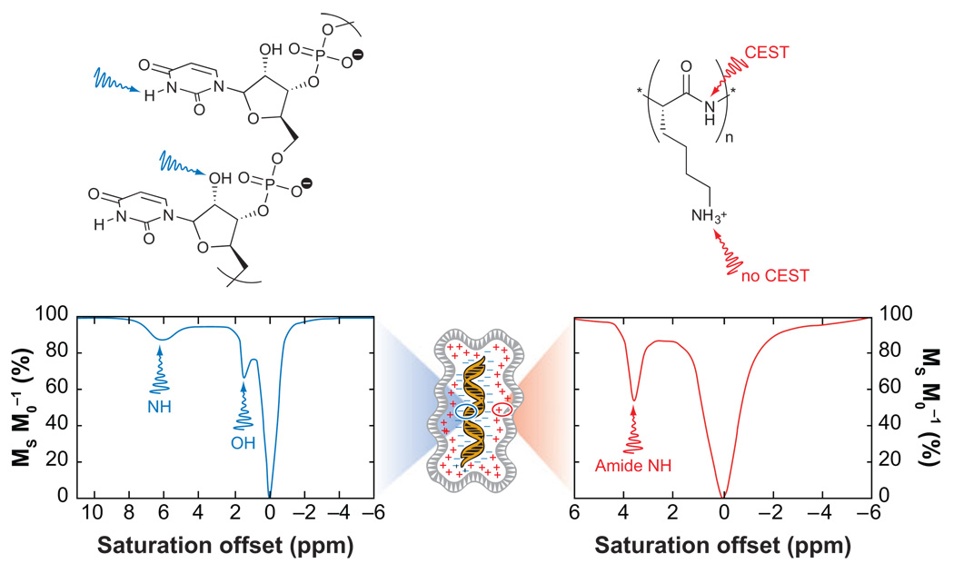
Magnetic resonance imaging (MRI) contrast agents have become an important tool in clinical medicine. The most common agents are Gd(3+)-based complexes that shorten bulk water T(1) by rapid exchange of a single inner-sphere water molecule with bulk solvent water. Current gadolinium agents lack tissue specificity and typically do not respond to their chemical environment. Recently, it has been demonstrated that MR contrast may be altered by an entirely different mechanism based on chemical exchange saturation transfer (CEST). CEST contrast can originate from exchange of endogenous amide or hydroxyl protons or from exchangeable sites on exogenous CEST agents. This has opened the door for the discovery of new classes of responsive agents ranging from MR gene reporter molecules to small molecules that sense their tissue environment and respond to biological events.


Figures

Figure 1
Figure 1
Schematic representations of the distribution of spins, aligned with and against the field (upper and lower energy levels, respectively) (above) and simulated NMR spectra (below) for two chemically distinct pools of nuclei (left), two spins after a saturation pulse has been applied to one pool (middle), and for a system undergoing chemical exchange after a saturation pulse has been applied to one pool (right).
Figure 2
Figure 2
Angiography by TOF-MRA is a form of saturation transfer experiment. An image slice, represented by the dotted line, is acquired by saturating the bulk water protons only in that image slice (left, the saturation is represented in blue). After a short time delay, the saturation of water protons in blood has been transferred out of the slice and replaced by unsaturated spins (middle). Thus, when the image is acquired, the stationary tissue appears dark but the blood is bright. In this way, angiograms may be acquired, such as the one of the carotid arteries shown (right).
Figure 3
Figure 3
An image of a mouse brain implanted with glioma cells expressing LRP (left hemisphere) and glioma cells (right hemisphere). On the left is an anatomical image, on the right a colorized difference image of the areas that exhibit CEST has been overlaid on the anatomical image. The LRP gives rise to a significant change in water signal intensity, allowing the genetically modified cells to be easily identified and tracked by CEST imaging. The area of CEST intensity around the skull is thought to be due to field inhomogeneities. Images reproduced, with permission, from Nature Biotechnology.
Figure 4
Figure 4
The structure of glycogen (top left) and its CEST spectra recorded at 200 and 400 MHz (top right). An anatomical image, with a presaturation pulse applied at +1 ppm, shows a darkening of the image due to the presence of glycogen (bottom left). Colorized CEST difference images acquired at 0, 20, 40, 60, 80, 100, and 120 min after administration of glucagon become progressively less orange, reflecting depletion of glycogen (bottom). MR images and CEST spectra reproduced from Proceedings of the National Academy of Sciences.
Figure 5
Figure 5
The setup of the phantom system used to image the CEST properties of hyperpolarized 129Xe (far left). Images of the phantom system showing the signal arising from free 129Xe (left) and free 129Xe after presaturation of the encapsulated xenon (right). The difference image (far right) shows that the CEST effect is only generated for those regions in which the avadin target is found. MR images from Reference 39. Reprinted with permission from AAAS.
Figure 6
Figure 6
The CEST spectra of barbituric acid (left) and a Eu3+ complex, EuDOTA-(glycine ethyl ester)4 (right). The CEST spectrum of barbituric acid is shown on the same scale as that of the Eu3+ complex (blue, right) for comparative purposes.
Figure 7
Figure 7
CEST imaging allows ratiometric determination of biologically relevant parameters such as pH and temperature. (Left) simulated CEST spectra of a cocktail of Eu3+ and Yb3+ DOTA-tetraamide complexes with varying pH. The insensitivity of CEST from the metal-bound water of the Eu3+ complex can be used as a concentration marker, whereas the pH-sensitive amide protons of the Yb3+ complexes can be used to measure pH. (Right) The CEST spectra of an Eu3+ DOTA-tetraamide complex at different temperatures.
Figure 8
Figure 8
Illustrating different methods of designing responsive PARACEST agents. (Left) The binding of glucose hinders water exchange, increasing CEST. (Middle) At physiological pH, binding of Zn2+ at its binding site above the Eu3+-OH2 molecule results in catalysis of proton exchange, altering the CEST properties. (Right) Altering the electronic effects of substituents can affect the interaction between the metal and water, which may be observed as a change in CEST.
Figure 9
Figure 9
Three examples of ways to enhance CEST sensitivity. (Left) A perfluorocarbon-filled nanoparticle has a large number of CEST agents incorporated onto the surface of the nanoparticle. (Middle) A spherical liposome is filled with a high concentration of TmDOTMA, a reagent that exchanges water rapidly and shifts the average position of the intraliposomal water downfield by nearly 4 ppm. (Right) An osmotically shrunk liposome filled with a high concentration of GdHP-DO3A, a clinically approved agent, results in a highly shifted intraliposomal water peak due to bulk susceptibility differences between the two water compartments.
Figure 10
Figure 10
A schematic representation of a gene therapy bundle of nucleic acids and cationic polymers. The RNA strand poly(rU) (left) has two sites that give rise to CEST. The amide protons in cationic polymers, such as PLL (right), can also give rise to a CEST effect.

References

    1. Caravan P, Ellison JJ, McMurry TJ, Lauffer RB. Gadolinium(III) chelates as MRI contrast agents: structure, dynamics, and applications. Chem. Rev. 1999;99:2293–1352. - PubMed
    1. Toth E, Merbach AE. Chemistry of Contrast Agents in Medical Magnetic Resonance Imaging. New York: Wiley; 2001.
    1. Kalman FK, Woods M, Caravan P, Jurek P, Spiller M, et al. Potentiometric and relaxometric properties of a gadolinium-based MRI contrast agent for sensing tissue pH. Inorg. Chem. 2007;46:5260–5270. - PMC - PubMed
    1. Aime S, Barge A, Botta M, Howard JAK, Kataky R, et al. Dependence of the relaxivity and luminescence of gadolinium and europium amino-acid complexes on hydrogencarbonate and pH. Chem. Commun. 1999;1999:1047–1048.
    1. Lowe MP, Parker D. Controllable pH modulation of lanthanide luminescence by intramolecular switching of the hydration state. Chem. Commun. 2000;2000:707–708.

Publication types

MeSH terms