. 2008 Jul 29;105(30):10338-43.
doi: 10.1073/pnas.0804659105. Epub 2008 Jul 22.

Mechanism for alternating access in neurotransmitter transporters

Affiliations

Mechanism for alternating access in neurotransmitter transporters

Lucy R Forrest et al. Proc Natl Acad Sci U S A. .

Abstract

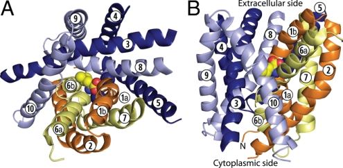
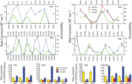
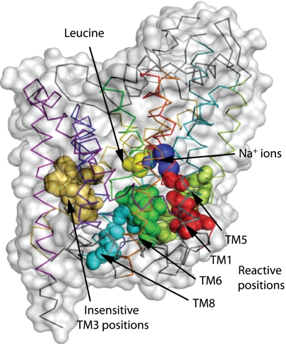
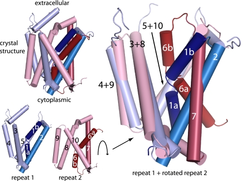
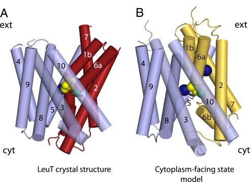
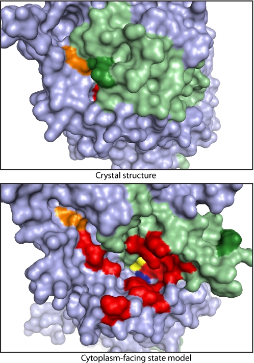
Crystal structures of LeuT, a bacterial homologue of mammalian neurotransmitter transporters, show a molecule of bound substrate that is essentially exposed to the extracellular space but occluded from the cytoplasm. Thus, there must exist an alternate conformation for LeuT in which the substrate is accessible to the cytoplasm and a corresponding mechanism that switches accessibility from one side of the membrane to the other. Here, we identify the cytoplasmic accessibility pathway of the alternate conformation in a mammalian serotonin transporter (SERT) (a member of the same transporter family as LeuT). We also propose a model for the cytoplasmic-facing state that exploits the internal pseudosymmetry observed in the crystal structure. LeuT contains two structurally similar repeats (TMs1-5 and TMs 6-10) that are inverted with respect to the plane of the membrane. The conformational differences between them result in the formation of the extracellular pathway. Our model for the cytoplasm-facing state exchanges the conformations of the two repeats and thus exposes the substrate and ion-binding sites to the cytoplasm. The conformational change that connects the two states primarily involves the tilting of a 4-helix bundle composed of transmembrane helices 1, 2, 6, and 7. Switching the tilt angle of this bundle is essentially equivalent to switching the conformation of the two repeats. Extensive mutagenesis of SERT and accessibility measurements, using cysteine reagents, are accommodated by our model. These observations may be of relevance to other transporter families, many of which contain internal inverted repeats.


Conflict of interest statement

The authors declare no conflict of interest.

Figures

Fig. 1.
Fig. 1.
Crystal structure of LeuT viewed from the extracellular side (A) and in the plane of the membrane (B). The leucine substrate and sodium ions are shown in spheres. Transmembrane domains from the first repeat (–5) are shown in darker colors (orange and dark blue), and those from the second repeat (–10) are in lighter colors (yellow and pale blue). The two structural repeats are divided up to form a bundle (yellow and orange) cradled within a scaffold (pale and dark blue).
Fig. 2.
Fig. 2.
Rate constants for reactivity of cytoplasmic pathway mutants with MTS reagents. (Upper) Calculated rate constants for inactivation of β-CIT binding to the indicated mutants by MTSEA (and MTSES for TMs 6 and 8). Bottom axes are SERT positions, and corresponding residues in LeuT are shown on the top axes (15). C and E indicate the cytoplasmic and extracellular ends, respectively, of each region. The green line indicates the accessibility in the cytoplasm-facing state model. The solvent-accessible surface area (SASA) for each residue was calculated as a percentage of the SASA of the same amino acid type (X) in a reference gly-X-gly tripeptide. From this, the equivalent percentage in the crystal structure was subtracted, so that the accessibility is positive only for residues that are exposed in the cytoplasm-facing state model; for all other residues, the accessibility is zero. Mutants with the highest rates of inactivation were further tested by measuring inactivation rates in the presence of 10 μM cocaine or ibogaine. (Lower) Rates of inactivation, relative to rates with the MTS reagent alone, for these selected positions. Error bars indicate the standard error of the means from three independent experiments. Asterisks indicate a significant difference from the control rate with MTS reagent alone (P < 0.05, paired Student's t test). TM5 data are from (20, 21).
Fig. 3.
Fig. 3.
Identification of the cytoplasmic permeation pathway in SERT. Residues in TM1 (red), -5 (lime), -6 (green), and -8 (cyan) of LeuT that correspond to those demonstrated by accessibility measurements (Fig. 2) to line the cytoplasmic pathway of SERT are shown as space-filling spheres. Five positions in TM3 with low accessibility (tan) are also shown. The C-alpha chain of LeuT (pdb code 2a65) is indicated by using the color coding of Yamashita et al. (9) along with a transparent representation of the protein surface.
Fig. 4.
Fig. 4.
Structural asymmetry of repeats in LeuT. TMs 1–10 of the LeuT crystal structure (Upper Left) were separated into the two structural repeats (Lower Left). TMs 1–2, dark blue; TMs 3–5, pale blue; TMs 6–7, dark red; TMs 8–10, pale red. The repeats were superimposed by aligning the positions of the last three helices in each repeat (Right). This required an ≈180° rotation of repeat 2 with respect to repeat 1 as calculated with SKA (45). Note the different orientation of the first two helices (particularly 1 and 6) in each repeat with respect to the other three helices.
Fig. 5.
Fig. 5.
Predicted conformational change in LeuT. The position of the four-helix bundle comprised of TMs 1–2 and 6–7 with respect to the scaffold (in blue) is shown along the plane of the membrane in red in the crystal structure (A) and in yellow in the model of the cytoplasm-facing state (B). The positions of the sodium ions (blue), the leucine substrate (yellow), and the Cα-atom of Ser-256 (green) are shown as spheres. For clarity, TM11 and -12 and the extracellular loops are not shown.
Fig. 6.
Fig. 6.
Molecular surface of LeuT, viewed from the cytoplasmic side of the membrane. (Upper) X-ray crystal structure. (Lower) Model of the cytoplasm-facing state. The bundle is shaded light green and the remainder of the protein light blue. The surface of leucine (yellow), Na2 (blue), the Arg-5 (green), and Asp-369 (orange) ion pair and residues corresponding to those found to be accessible in SERT (red and orange) are highlighted.

References

    1. Jardetzky O. Simple allosteric model for membrane pumps. Nature. 1966;211:969–970. - PubMed
    1. Takahashi M, Kondou Y, Toyoshima C. Interdomain communication in calcium pump as revealed in the crystal structures with transmembrane inhibitors. Proc Natl Acad Sci USA. 2007;104:5800–5805. - PMC - PubMed
    1. Olesen C, et al. The structural basis of calcium transport by the calcium pump. Nature. 2007;450:1036–1042. - PubMed
    1. Hirai T, et al. Three-dimensional structure of a bacterial oxalate transporter. Nat Struct Biol. 2002;9:597–600. 2002. - PubMed
    1. Abramson J, et al. Structure and mechanism of the lactose permease of Escherichia coli. Science. 2003;301:610–615. - PubMed

Publication types

LinkOut - more resources