. 2008 Oct;18(10):806-17.
doi: 10.1093/glycob/cwn070. Epub 2008 Jul 22.

Analysis of glycosyltransferase expression in metastatic prostate cancer cells capable of rolling activity on microvascular endothelial (E)-selectin

Affiliations

Analysis of glycosyltransferase expression in metastatic prostate cancer cells capable of rolling activity on microvascular endothelial (E)-selectin

Steven R Barthel et al. Glycobiology. 2008 Oct.

Abstract

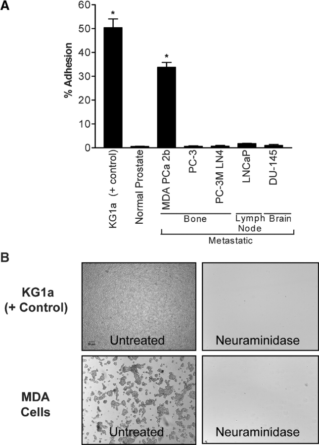
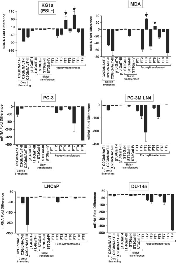
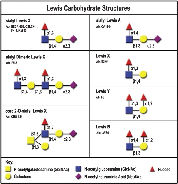
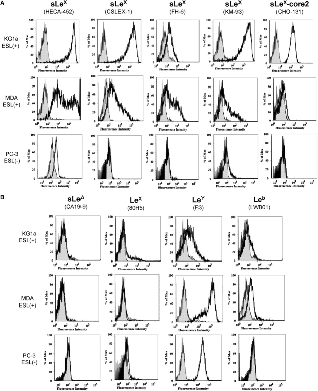
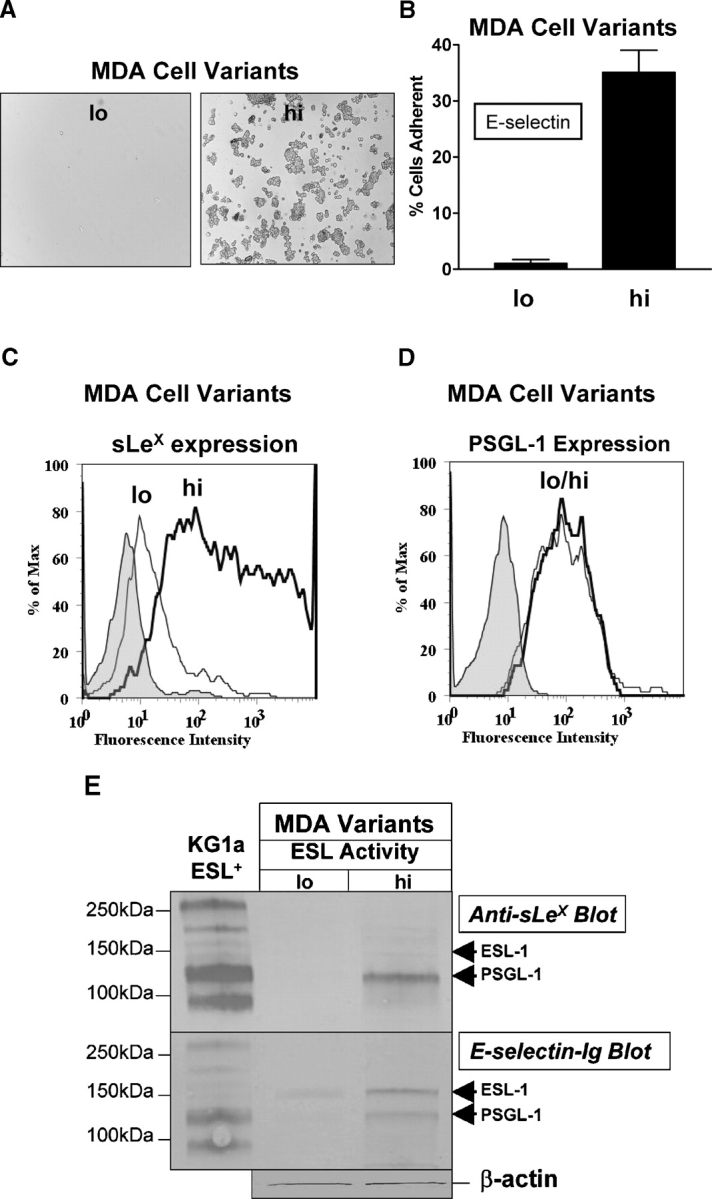
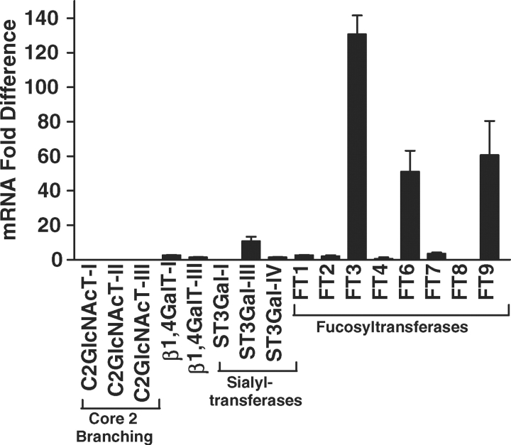
Prostate cancer (PCa) cell tethering and rolling on microvascular endothelium has been proposed to promote the extravasation of PCa cells. We have shown that these adhesive events are mediated through binding interactions between endothelial (E)-selectin and Lewis carbohydrates on PCa cells. Prior data indicate that E-selectin-mediated rolling of bone-metastatic PCa MDA PCa 2b (MDA) cells is dependent on sialyl Lewis X (sLe(X))-bearing glycoproteins. To explore the molecular basis of sLe(X) synthesis and E-selectin ligand (ESL) activity on PCa cells, we compared and contrasted the expression level of glycosyltransferases, characteristically involved in sLe(X) and ESL synthesis, in ESL(+) MDA cells among other ESL(-) metastatic PCa cell lines. We also created and examined ESL(hi) and ESL(lo) variants of MDA cells to provide a direct comparison of the glycosyltransferase expression level. We found that normal prostate tissue and all metastatic PCa cell lines expressed glycosyltransferases required for sialo-lactosamine synthesis, including N-acetylglucosaminyl-, galactosyl-, and sialyltransferases. However, compared with expression in normal prostate tissue, ESL(+) MDA cells expressed a 31- and 10-fold higher level of alpha1,3 fucosyltransferases (FT) 3 and 6, respectively. Moreover, FT3 and FT6 were expressed at 2- to 354-fold lower levels in ESL(-) PCa cell lines. Consistent with these findings, ESL(hi) MDA cells expressed a 131- and 51-fold higher level of FT3 and FT6, respectively, compared with expression in ESL(lo) MDA cells. We also noted that alpha1,3 FT7 was expressed at a 5-fold greater level in ESL(hi) MDA cells. Furthermore, ESL(lo) MDA cells did not display sLe(X) on glycoproteins capable of bearing sLe(X), notably P-selectin glycoprotein ligand-1. These results implicate the importance of alpha1,3 FT3, FT6, and/or FT7 in sLe(X) and ESL synthesis on metastatic PCa cells.


Figures

Fig. 1
Fig. 1
ESL activity of metastatic PCa MDA cells is dependent on sialylated membrane structures. Metastatic PCa cell lines, MDA, PC-3, PC-3M LN4, LNCaP, and DU-145, normal prostate epithelia, and control ESL+ KG1a cells were examined for ESL activity in a microtiter 96-well E-selectin-Ig-binding assay. (A) KG1a and MDA cells exhibited ESL activity, while all other cells had no activity. Negative control wells, consisting of cells plated on human IgG-adsorbed plastic or cells incubated in an EDTA-containing assay medium, had no ESL activity (not shown). (B) Pretreatment of MDA and KG1a cells with Vibrio cholerae neuraminidase completely ablated ESL activity. Experiments were performed in triplicate wells on at least three separate occasions. *P < 0.01, Statistical significant difference compared with adhesion to human IgG control; one-way ANOVA with Dunnett's post-test.
Fig. 2
Fig. 2
Elevated α1,3 fucosyltransferase expression is related to ESL activity on metastatic PCa MDA cells. Real-time PCR analysis of putative sLeX-synthesizing glycosyltransferases, including N-acetylglucosaminyl-, galactosyl-, sialyl-, and fucosyltransferases, was performed on cDNA isolated from control ESL+ KG1a cells, metastatic PCa cell lines, MDA, PC-3, PC-3M LN4, LNCaP, and DU-145, and normal prostate tissue. Expression is reported as the mean (SEM) mRNA fold difference after normalizing to expression level in fresh normal prostate epithelial tissue. Arrows denote elevation of FT3 and FT6 expression in ESL+ MDA cells and FT4 and FT7 in ESL+ KG1a cells. Experiments were performed a minimum of three times.
Fig. 3
Fig. 3
Structures of Lewis carbohydrates. Schematic of Lewis carbohydrate determinants recognized by respective mAbs in flow cytometric assays. Epitopes recognized by HECA-452, CSLEX-1, FH-6, KM-93, CA19-9, and F3 (Kannagi and Hakomori 2001) as well as CHO-131 (Walcheck et al. 2002), 80H5 (Benharroch et al. 2000), and LWB01 (Torrado et al. 1992) have been reported.
Fig. 4
Fig. 4
SLeX and not sLea moieties are elevated on ESL+ metastatic PCa MDA cells. (A) Flow cytometric analysis of sLeX moieties and (B) sLea, LeX, Ley, and Leb was performed on control ESL+ KG1a cells as well as on ESL+ MDA and ESL PC-3 cells. Open histograms: staining with anti-Lewis carbohydrate mAb; shaded histograms: staining with isotype control mAb. ESL+ cells were recognized by anti-sLeX mAbs, HECA-452, CSLEX-1, FH-6, KM-93, whereas ESL PC-3 cells were negative for sLeX. ESL+ and ESL PCa cells were negative for sLea and positive for LeY. These experiments were performed a minimum of three times.
Fig. 5
Fig. 5
ESLhi metastatic PCa MDA cell variants express sLeX- and E-selectin-Ig-reactive PSGL-1. (A and B) MDA cell variants were separated into ESLlo and ESLhi based on their E-selectin-binding characteristics. (C and D) Flow cytometric analysis of expression of sLeX and PSGL-1 on ESLlo (thin-lined histogram) and ESLhi (thick-lined histogram) MDA cell variants. Expression of sLeX was assayed with mAb HECA-452 and control rat IgM isotype (shaded histogram). Expression of PSGL-1 was assayed with mAb PL2 and control mouse IgG1 isotype (shaded histogram). Histograms are representative of experiments performed on at least five different MDA cell variants. (E) Western blot analysis of sLeX and ESL was performed on ESLlo and ESLhi MDA cell variants. Lysates from control ESL+ HPC (KG1a) cells and ESLlo and ESLhi variants were subjected to SDS–PAGE on 4–20% reducing gels, transferred to immunoblot membrane, and blotted with anti-sLeX mAb HECA-452 or with E-selectin-Ig chimera. There were no glycoproteins bearing sLeX on ESLlo MDA cell variants, while sLeX and E-selectin-binding determinants were expressed by PSGL-1 on ESLhi MDA cell variants. Blotting with secondary rat IgM alone or with E-selectin-Ig in 5 mM EDTA did not result in any staining activity. These experiments were performed a minimum of five times.
Fig. 6
Fig. 6
α2,3 Sialyltransferase III and α1,3 FT3, FT6, FT7, and FT9 are elevated in ESLhi metastatic PCa MDA cell variants. Real-time PCR analysis of N-acetylglucosaminyl-, galactosyl-, sialyl-, and fucosyltransferases was performed on cDNA isolated from ESLlo and ESLhi metastatic PCa MDA cell variants. Expression in ESLhi MDA cells is reported as the mean (SEM) fold difference after normalizing to expression levels in ESLlo MDA cells. ST3Gal-III and FT3, FT6, FT7, and FT9 were elevated in ESLhi MDA cells. Experiments were performed a minimum of three times.

Similar articles

Cited by

References

    1. Aubert M, Panicot-Dubois L, Crotte C, Sbarra V, Lombardo D, Sadoulet MO, Mas E. Peritoneal colonization by human pancreatic cancer cells is inhibited by antisense FUT3 sequence. Int J Cancer. 2000;88:558–565. - PubMed
    1. Barthel SR, Gavino JD, Descheny L, Dimitroff CJ. Targeting selectins and selectin ligands in inflammation and cancer. Expert Opin Ther Targets. 2007;11:1473–1491. - PMC - PubMed
    1. Benharroch D, Dima E, Levy A, Ohana-Malka O, Ariad S, Prinsloo I, Mejirovsky E, Sacks M, Gopas J. Differential expression of sialyl and non-sialyl-CD15 antigens on Hodgkin-Reed-Sternberg cells: Significance in Hodgkin's disease. Leuk Lymphoma. 2000;39:185–194. - PubMed
    1. Benjamin R. Neurologic complications of prostate cancer. Am Fam Physician. 2002;65:1834–1840. - PubMed
    1. Biol-N’garagba MC, Niepceron E, Mathian B, Louisot P. Glucocorticoid-induced maturation of glycoprotein galactosylation and fucosylation processes in the rat small intestine. J Steroid Biochem Mol Biol. 2003;84:411–422. - PubMed

Publication types