. 2008 Aug;67(8):773-83.
doi: 10.1097/NEN.0b013e318180ec47.

Relationships between expression of apolipoprotein E and beta-amyloid precursor protein are altered in proximity to Alzheimer beta-amyloid plaques: potential explanations from cell culture studies

Affiliations

Relationships between expression of apolipoprotein E and beta-amyloid precursor protein are altered in proximity to Alzheimer beta-amyloid plaques: potential explanations from cell culture studies

Steven W Barger et al. J Neuropathol Exp Neurol. 2008 Aug.

Abstract

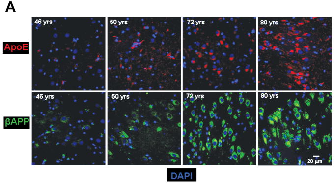
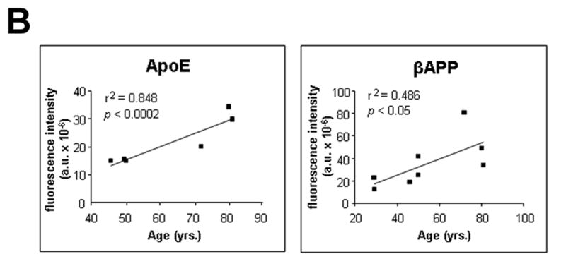
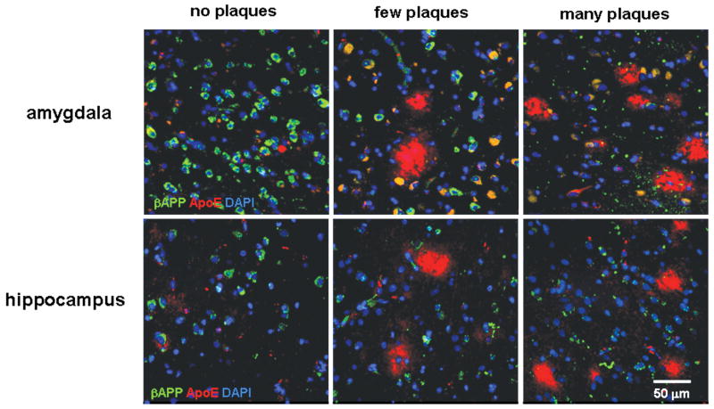
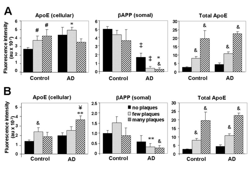
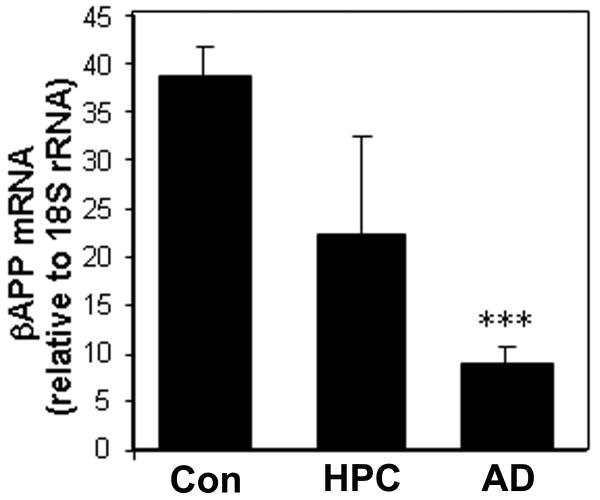
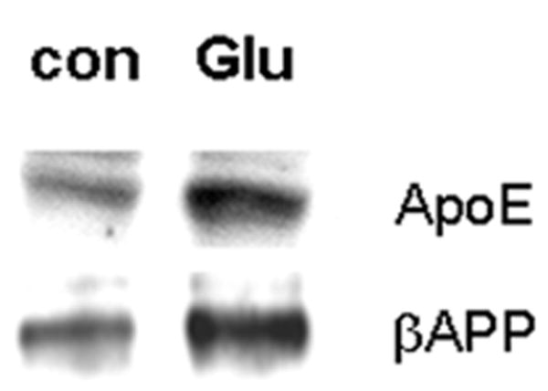
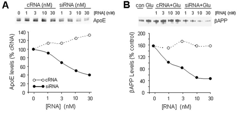
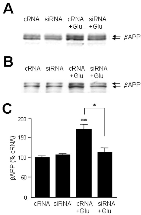
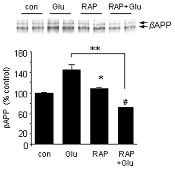
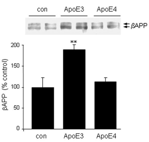
Theories regarding the initiation and progression of Alzheimer disease (AD) often consider potential roles played by elevations of beta-amyloid precursor protein (betaAPP). Because it is the source of amyloid beta-peptide, betaAPP may simply contribute more pathogenic stimulus when elevated; some analyses have, however, reported a decline in betaAPP in AD. We found a progressive increase in neuronal betaAPP expression with increasing age in the brains of nondemented individuals, whereas in AD patient samples, betaAPP antigenicity decreased in neuronal somata in a manner that correlated with accumulation of mature amyloid beta-peptide plaques. In contrast, apolipoprotein E (ApoE) expression correlated with accumulation of plaques, and even greater amounts of ApoE were detected in plaques. Induction of betaAPP by glutamate in neuronal cell cultures was found to depend upon ApoE levels or activity. Thus, elevations in expression of ApoE and betaAPP by cellular stresses are likely normally linked in vivo, and uncoupling of this link, or other pathologic events in AD initiation, may leave neurons with diminished betaAPP expression, which might in turn reduce their resistance to stressors.


Figures

Figure 1
Figure 1
Age-related changes in expression of βAPP and ApoE. (A) βAPP (green) and ApoE (red) were detected by immunofluorescence in tissue sections from hippocampus at the level of the lateral geniculate nucleus. Results are shown for 4 non-demented individuals across an age span of 46 to 80 years. Blue represents DAPI staining of cellular DNA. Images were digitized at 20× magnification. Scale bar represents 20 μm. (B) Quantitation of βAPP and ApoE immunofluorescence intensity was obtained by thresholding βAPP grayscale images and integrating pixels as described in Materials and Methods. Values reflect the mean of 3 images per hippocampus of 6 (ApoE) or 8 (βAPP) non-demented individuals.
Figure 1
Figure 1
Age-related changes in expression of βAPP and ApoE. (A) βAPP (green) and ApoE (red) were detected by immunofluorescence in tissue sections from hippocampus at the level of the lateral geniculate nucleus. Results are shown for 4 non-demented individuals across an age span of 46 to 80 years. Blue represents DAPI staining of cellular DNA. Images were digitized at 20× magnification. Scale bar represents 20 μm. (B) Quantitation of βAPP and ApoE immunofluorescence intensity was obtained by thresholding βAPP grayscale images and integrating pixels as described in Materials and Methods. Values reflect the mean of 3 images per hippocampus of 6 (ApoE) or 8 (βAPP) non-demented individuals.
Figure 2
Figure 2
βAPP levels decrease and ApoE levels increase with advancing AD pathology. βAPP (green) and ApoE (red) were detected in areas of amygdala or hippocampus with no Aβ deposits, a few Aβ plaques, or multiple Aβ plaques. Colocalization of βAPP and ApoE in neurons appears yellow-green. Blue is DAPI staining of cellular DNA. Note the increased βAPP-positive dystrophic neurites in the panels with many plaques. The scale bar represents 50 μm.
Figure 3
Figure 3
βAPP and ApoE are differentially modulated in relation to plaque pathology. Intensities of βAPP fluorescence in neuronal somata, ApoE fluorescence in any cellular compartment, and total ApoE fluorescence were determined using analyses described in Materials and Methods. Fields were grouped according to “no plaques” (0), “few plaques” (–4), or “many plaques” (≥5), in amygdala (A) or hippocampus (B). Values reflect the mean ± SEM from 13 (no plaques), 12 (few plaques), or 7 (many plaques) individuals. & p < 0.02, # p < 0.05 vs. control, no plaques; p < 0.001, ** p < 0.01, * p < 0.05 vs. corresponding control value; ¥ p < 0.01 vs. AD, no plaques.
Figure 4
Figure 4
βAPP mRNA declines in AD initiation and progression. Total RNA was extracted from amygdala of brains diagnosed as control, AD (n = 5), or high-pathology controls (HPC; clinically nondemented but with AD pathology; n = 3). RNA was subjected to qRT-PCR and scaled relative to input RNA quantity in a standard curve. Final values were corrected by the signal for 18S rRNA. ***p < 0.00006 vs. control (Student t-test)
Figure 5
Figure 5
Glutamate elevates ApoE and βAPP levels coordinately in primary neuronal cultures. Primary cortical neurons were left untreated or exposed to 20 μM glutamate for 20 hours, and then βAPP and ApoE were assessed by Western blot analysis. Data are representative of >10 experiments.
Figure 6
Figure 6
Glutamate-induced elevation of βAPP is dependent upon ApoE expression. (A) The human neuronal cell line NTera2 was treated for 48 hours with the indicated concentrations of control RNA (“cRNA”; open circles) or siRNA directed against ApoE (filled circles). ApoE levels were assayed by Western blot analysis to verify effectiveness of siRNA. (B) NTera2 cells were treated with ApoE siRNA (filled circles), cRNA (open circles) or no RNA (lanes 1 and 2). After 48 hours, glutamate (20 μM) was applied to all but one set of cultures (lane 1). After 20 hours, βAPP levels were assayed by Western blot analysis. In both A and B, 1-way ANOVA indicates significant differences between the control and siRNA curves (p < 0.008).
Figure 7
Figure 7
Glutamate→ApoE→βAPP axis manifest in neuronal cultures. Cultures of primary rat cortical neurons (A) or NTera2 cells (B) were treated for 48 hours with 30 nM cRNA or siRNA directed against ApoE. Glutamate (20 μM) was then applied for 20 hours, after which βAPP levels were assayed by Western blot analysis. (C) Results from 3 experiments with NTera2 cells were quantified; *p < 0.05, siRNA+Glu vs. cRNA+Glu, **p < 0.01 cRNA+Glu vs. cRNA alone.
Figure 8
Figure 8
LRP-ApoE interactions modulate glutamate induction of βAPP. NTera2 cells were treated with 20 μM glutamate in the presence or absence of 200 nM receptor-associated protein (RAP); additional cultures were treated with RAP alone. βAPP expression was determined 20 hours later by Western blot analysis. *p < 0.05; **p < 0.01; #p < 0.05 (vs. RAP alone).
Figure 9
Figure 9
ApoE directly elevates βAPP expression in an isoform-specific manner. Mouse primary cortical neurons were treated for 20 hours with 5 μg/ml ApoE3 or ApoE4. βAPP levels were then assessed by Western blot analysis. Values represent the mean ± SEM of triplicate determinations. **p < 0.01 vs. control or ApoE4.

References

    1. Rosen DR, Martin-Morris L, Luo LQ, et al. A Drosophila gene encoding a protein resembling the human β-amyloid protein precursor. Proc Natl Acad Sci U S A. 1989;86:2478–82. - PMC - PubMed
    1. Dawson GR, Seabrook GR, Zheng H, et al. Age-related cognitive deficits, impaired long-term potentiation and reduction in synaptic marker density in mice lacking the beta-amyloid precursor protein. Neuroscience. 1999;90:1–13. - PubMed
    1. Masliah E, Westland CE, Rockenstein EM, et al. Amyloid precursor proteins protect neurons of transgenic mice against acute and chronic excitotoxic injuries in vivo. Neuroscience. 1997;78:135–46. - PubMed
    1. Prasher VP, Farrer MJ, Kessling AM, et al. Molecular mapping of Alzheimer-type dementia in Down's syndrome. Ann Neurol. 1998;43:380–83. - PubMed
    1. Griffin WS, Sheng JG, McKenzie JE, et al. Life-long overexpression of S100beta in Down's syndrome: implications for Alzheimer pathogenesis. Neurobiol Aging. 1998;19:401–405. - PMC - PubMed

Publication types

MeSH terms