

Substantial alterations of the cutaneous bacterial biota in psoriatic lesions
- PMID: 18648509
- PMCID: PMC2447873
- DOI: 10.1371/journal.pone.0002719
Substantial alterations of the cutaneous bacterial biota in psoriatic lesions
Abstract
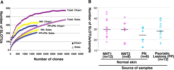
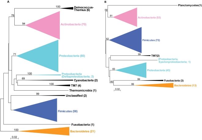
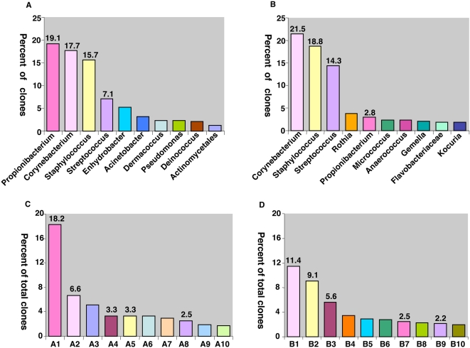
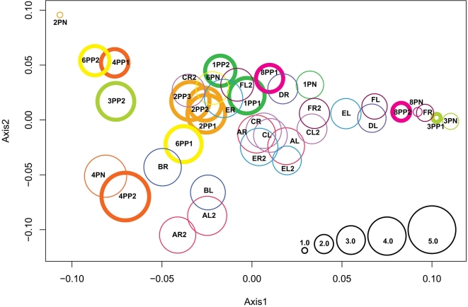
For psoriasis, an idiopathic inflammatory disorder of the skin, the microbial biota has not been defined using cultivation-independent methods. We used broad-range 16S rDNA PCR for archaea and bacteria to examine the microbiota of normal and psoriatic skin. From 6 patients, 19 cutaneous samples (13 from diseased skin and 6 from normal skin) were obtained. Extracted DNA was subjected to the broad range PCR, and 1,925 cloned products were compared with 2,038 products previously reported from healthy persons. Using 98% sequence identity as a species boundary, 1,841 (95.6%) clones were similar to known bacterial 16S rDNA, representing 6 phyla, 86 genera, or 189 species-level operational taxonomic unit (SLOTU); 84 (4.4%) clones with <98% identity probably represented novel species. The most abundant and diverse phylum populating the psoriatic lesions was Firmicutes (46.2%), significantly (P<0.001) overrepresented, compared to the samples from uninvolved skin of the patients (39.0%) and healthy persons (24.4%). In contrast, Actinobacteria, the most prevalent and diverse phylum in normal skin samples from both healthy persons (47.6%) and the patients (47.8%), was significantly (P<0.01) underrepresented in the psoriatic lesion samples (37.3%). Representation of Propionibacterium species were lower in the psoriatic lesions (2.9+/-5.5%) than from normal persons (21.1+/-18.2%; P<0.001), whereas normal skin from the psoriatic patients showed intermediate levels (12.3+/-21.6%). We conclude that psoriasis is associated with substantial alteration in the composition and representation of the cutaneous bacterial biota.
Conflict of interest statement
Figures
Similar articles
-
Bacterial biota in the human distal esophagus.Proc Natl Acad Sci U S A. 2004 Mar 23;101(12):4250-5. doi: 10.1073/pnas.0306398101. Epub 2004 Mar 11. Proc Natl Acad Sci U S A. 2004. PMID: 15016918 Free PMC article.
-
Alteration of the cutaneous microbiome in psoriasis and potential role in Th17 polarization.Microbiome. 2018 Sep 5;6(1):154. doi: 10.1186/s40168-018-0533-1. Microbiome. 2018. PMID: 30185226 Free PMC article.
-
Psoriatic lesions are characterized by higher bacterial load and imbalance between Cutibacterium and Corynebacterium.J Am Acad Dermatol. 2020 Apr;82(4):955-961. doi: 10.1016/j.jaad.2019.06.024. Epub 2019 Jun 19. J Am Acad Dermatol. 2020. PMID: 31228520
-
The Microbiome in Psoriasis and Psoriatic Arthritis: The Skin Perspective.J Rheumatol Suppl. 2018 Jun;94:30-31. doi: 10.3899/jrheum.180133. J Rheumatol Suppl. 2018. PMID: 29858350 Review.
-
FEATURES OF MICROBIOTA IN PSORIATIC DISEASE: FROM SKIN AND GUT PERSPECTIVES (REVIEW).Georgian Med News. 2019 Feb;(287):98-104. Georgian Med News. 2019. PMID: 30958298 Review.
Cited by
-
The Contribution of the Skin Microbiome to Psoriasis Pathogenesis and Its Implications for Therapeutic Strategies.Medicina (Kaunas). 2024 Oct 3;60(10):1619. doi: 10.3390/medicina60101619. Medicina (Kaunas). 2024. PMID: 39459406 Free PMC article. Review.
-
Exploratory multi-omics analysis reveals host-microbe interactions associated with disease severity in psoriatic skin.EBioMedicine. 2024 Jul;105:105222. doi: 10.1016/j.ebiom.2024.105222. Epub 2024 Jun 25. EBioMedicine. 2024. PMID: 38924840 Free PMC article.
-
Microbiome, Autoimmune Diseases and HIV Infection: Friends or Foes?Nutrients. 2019 Nov 2;11(11):2629. doi: 10.3390/nu11112629. Nutrients. 2019. PMID: 31684052 Free PMC article. Review.
-
Unveiled feather microcosm: feather microbiota of passerine birds is closely associated with host species identity and bacteriocin-producing bacteria.ISME J. 2019 Sep;13(9):2363-2376. doi: 10.1038/s41396-019-0438-4. Epub 2019 May 24. ISME J. 2019. PMID: 31127178 Free PMC article.
-
Characterization and Analysis of the Skin Microbiota in Rosacea: Impact of Systemic Antibiotics.J Clin Med. 2020 Jan 9;9(1):185. doi: 10.3390/jcm9010185. J Clin Med. 2020. PMID: 31936625 Free PMC article.
References
-
- Fredricks DN. Microbial ecology of human skin in health and disease. J.Investig.Dermatol.Symp.Proc. 2001;6:167–169. - PubMed
Publication types
MeSH terms
Substances
Grants and funding
LinkOut - more resources
Full Text Sources
Other Literature Sources
Medical
Molecular Biology Databases
