. 2008 May;15(1):119-32.
doi: 10.1080/15419060802013885.

Trafficking dynamics of glycosylated pannexin 1 proteins

Affiliations

Trafficking dynamics of glycosylated pannexin 1 proteins

Daniela Boassa et al. Cell Commun Adhes. 2008 May.

Abstract

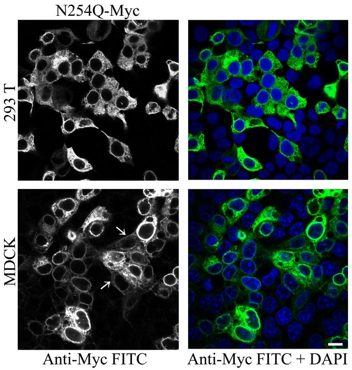
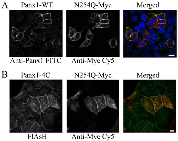
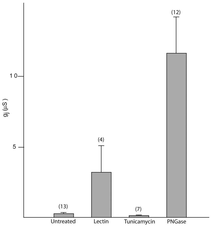
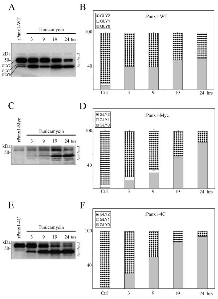
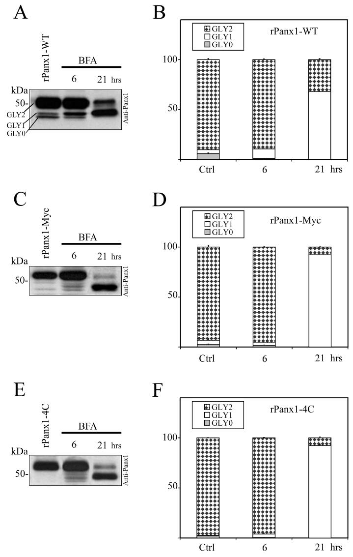
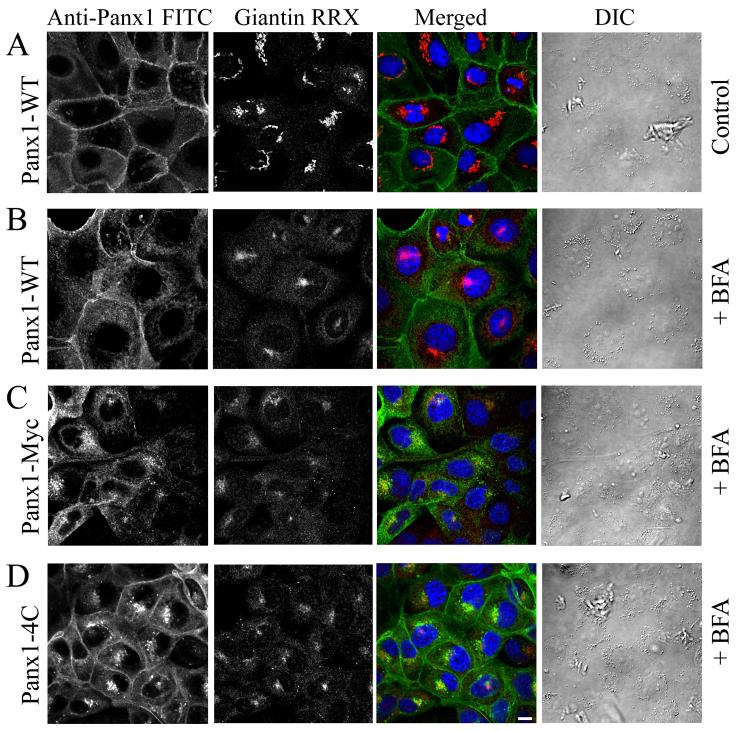
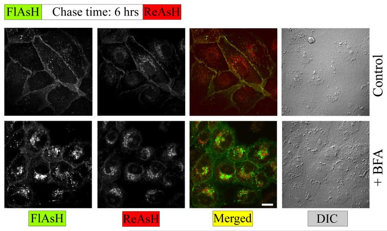
Pannexins are mammalian orthologs of innexins and have a predicted topological folding pattern similar to that of connexins, except they are glycosylated. Rat pannexin 1 is glycosylated at N254 and this residue is important for plasma membrane targeting. Here we demonstrate that cell surface expression levels of the rat pannexin 1 N254Q mutant are rescued by coexpression with the wild-type protein. In paired Xenopus oocytes, the functional effect of this rescue is inconsequential; however, cell surface deglycosylation by PNGase F significantly enhanced functional gap junction formation. In mammalian cells, wild-type oligomers traffic at a slower rate than Myc-or tetracysteine domain-tagged versions, a behavior opposite to that of tagged connexins. The temporal differences of Panx1 trafficking correlate with spatial differences of intracellular localizations induced by Golgi blockage by Brefeldin-A or glycosylation prevention by tunicamycin. Therefore, Panx1 has kinetics and dynamics that make it unique to serve distinct functions separate from connexin-based channels.


Figures

Figure 1
Figure 1. Preventing N-glycosylation at the N254 residue induces high ER retention and inhibits cell surface expression
Immunolabeling of HEK-293T and MDCK cells transiently expressing Myc-tagged N254Q mutant rPanx1 proteins. Anti-Myc staining (left column in black and white) and the merged view with a nuclear stain DAPI (right column) are shown, and indicate high efficiency of transfection. White arrows show staining of the mutants localized at the cell surface in MDCK cells. The scale bar equals 10 μm.
Figure 2
Figure 2. Cell surface expression of the rPanx1 N254Q mutant is rescued by co-expression with rPanx1-wild type proteins
(A) MDCK cells transiently co-expressing rPanx1-WT and Myc-tagged N254Q mutant proteins were stained with anti-Panx1 (FITC, left side in black and white), and anti-Myc (CY5, middle in black and white). Cell surface expression of the mutant proteins was rescued as shown by the yellow color in the merged view (right side, green for Anti-Panx1, red for anti-Myc and blue for nuclear stain DAPI). (B) MDCK cells stably expressing Panx1-4C and transiently transfected with Myc-tagged N254Q mutant Panx1 were stained with green profluorescent FlAsH-EDT2 (left side in black and white) and anti-Myc (CY5, middle in black and white). The cell surface expression of the mutant protein was also rescued by co-expression with the 4C-tagged Panx1 protein as displayed by the yellow in the merged view (FlAsH-EDT2 in green and anti-Myc in red). The scale bars equal 10 μm.
Figure 3
Figure 3. Effect of deglycosylation treatments on the junctional conductance of oocyte pairs expressing mPanx1
Panx1-WT cRNA was injected 2-3 days before pairing. The oocytes were treated with lectin (glycine max, 10 μg/ml), tunicamycin (1 μg/ml), or PNGase F (10 units/ml) for 30 minutes before pairing. Junctional conductance was determined 6 hours after pairing. Treatments that promoted the removal of carbohydrate groups from the extracellular surface significantly increased junctional conductance.
Figure 4
Figure 4. Comparison of the effect of glycosylation inhibition on rPanx1-WT, rPanx1-Myc and rPanx1-4C tagged proteins
Western blot analysis of cellular lysates from MDCK cells stably expressing rPanx1-WT (A), rPanx1-Myc (C), or rPanx1-4C (E) before and after tunicamycin treatment (2 μg/ml) for the indicated time periods (hrs). The blots were hybridized with anti-Panx1 antibody. The relative densities of the different forms of the protein (GLY0, non-glycosylated core protein, GLY1, high mannose type glycoprotein, and GLY2, fully processed glycoprotein) were quantified (y axis, normalized value) and represent the mean ± S.E. of three independent Western blots respectively for rPanx1-WT (B), rPanx1-Myc (D), or rPanx1-4C (F). In these samples under control conditions we observed some variations in GLY0 and GLY1 bands in tagged Panx1 while the majority of the proteins (more than 90%) were found in the GLY2 form. Glycosylation inhibition over periods of time induced a significant increase in the GLY0, and a decrease in the GLY2 species for both rPanx1-WT and tagged proteins. However, comparison of the trafficking kinetics of tagged rPanx1 versus WT revealed that WT GLY2 oligomers appear to be degraded at a slower rate than Myc or tetracysteine domain tagged versions.
Figure 5
Figure 5. Comparison of the effect of BFA treatment on rPanx1-WT, rPanx1-Myc and rPanx1-4C tagged proteins
Western blot analysis of cellular lysates from MDCK cells stably expressing rPanx1-WT (A), rPanx1-Myc (C), or rPanx1-4C (E) before and after BFA treatment (5 μg/ml) for the indicated time periods (hrs). The blots were hybridized with anti-Panx1 antibody and the relative densities (in the y axis as normalized values) of the different forms of protein analyzed as in Figure 3. Summary from three independent Western blots (mean ± S.E.) respectively for rPanx1-WT (B), rPanx1-Myc (D), or rPanx1-4C (F) is reported. A significant decrease in GLY2 and increase in GLY1 was observed with longer incubation time only, and to a greater extent for the tagged rPanx1 versus the rPanx1-WT.
Figure 6
Figure 6. Cell surface expression of tagged rPanx1 is affected more than rPanx1-WT by long incubation time with BFA
(A) MDCK cells stably expressing rPanx1-WT in control conditions (not treated with BFA). To block protein secretion MDCK cells stably expressing rPanx1-WT (B), rPanx1-Myc (C) or rPanx1-4C (D) tagged proteins were treated with 5 μg/ml BFA for 21 hours. Confocal images of immunolabeled cells stained for anti-Panx1 (FITC) and with antibodies for the Golgi protein giantin (RRX) are displayed. The Golgi marker serves as positive control for the effectiveness of the drug in causing and maintaining Golgi fragmentation. Differential interference contrast (DIC) images show that cells morphology and viability was unaffected by the treatment. Cell surface expression is more prominent for rPanx1-WT, however the intracellular localization of newly synthesized proteins is predominant for the rPanx1-Myc and rPanx1-4C. The scale bar equals 10 μm.
Figure 7
Figure 7. Pulse chase experiments using FlAsH and ReAsH reveals temporal and spatial segregation of different pools of rPanx1-4C following BFA treatment
MDCK cells stably expressing rPanx1-4C were labeled with green profluorescent FlAsH-EDT2, incubated for 6 hrs in regular medium (control) or in the presence of 5 μg/ml BFA, stained with red profluorescent ReAsH-EDT2, fixed, and imaged. After BFA treatment the nascent rPanx1-4C proteins (labeled with ReAsH) are accumulated within intracellular compartments. The older proteins (labeled with FlasH) are partially internalized although the majority is still localized at the cell surface. The scale bar equals 10 μm.

References

    1. Bao L, Locovei S, Dahl G. Pannexin membrane channels are mechanosensitive conduits for ATP. FEBS Lett. 2004;572:65–68. - PubMed
    1. Bao L, Samuels S, Locovei S, Macagno ER, Muller KJ, Dahl G. Innexins form two types of channels. FEBS Lett. 2007;581:5703–5708. - PMC - PubMed
    1. Boassa D, Ambrosi C, Qiu F, Dahl G, Gaietta G, Sosinsky G. Pannexin1 channels contain a glycosylation site that targets the hexamer to the plasma membrane. J Biol Chem. 2007;282:31733–31743. - PubMed
    1. Bruzzone R, Hormuzdi SG, Barbe MT, Herb A, Monyer H. Pannexins, a family of gap junction proteins expressed in brain. Proc Natl Acad Sci U S A. 2003;100:13644–13649. - PMC - PubMed
    1. Bukauskas FF, Jordan K, Bukauskiene A, Bennett MV, Lampe PD, Laird DW, Verselis VK. Clustering of connexin 43-enhanced green fluorescent protein gap junction channels and functional coupling in living cells. Proc Natl Acad Sci U S A. 2000;97:2556–2561. - PMC - PubMed

Publication types

LinkOut - more resources