. 2008 Jul 23;28(30):7458-66.
doi: 10.1523/JNEUROSCI.0197-08.2008.

Ca2+-dependent, phospholipid-binding residues of synaptotagmin are critical for excitation-secretion coupling in vivo

Affiliations

Ca2+-dependent, phospholipid-binding residues of synaptotagmin are critical for excitation-secretion coupling in vivo

Brie E Paddock et al. J Neurosci. .

Abstract

Synaptotagmin I is the Ca(2+) sensor for fast, synchronous release of neurotransmitter; however, the molecular interactions that couple Ca(2+) binding to membrane fusion remain unclear. The structure of synaptotagmin is dominated by two C(2) domains that interact with negatively charged membranes after binding Ca(2+). In vitro work has implicated a conserved basic residue at the tip of loop 3 of the Ca(2+)-binding pocket in both C(2) domains in coordinating this electrostatic interaction with anionic membranes. Although results from cultured cells suggest that the basic residue of the C(2)A domain is functionally significant, such studies provide contradictory results regarding the importance of the C(2)B basic residue during vesicle fusion. To directly test the functional significance of each of these residues at an intact synapse in vivo, we neutralized either the C(2)A or the C(2)B basic residue and assessed synaptic transmission at the Drosophila neuromuscular junction. The conserved basic residues at the tip of the Ca(2+)-binding pocket of both the C(2)A and C(2)B domains mediate Ca(2+)-dependent interactions with anionic membranes and are required for efficient evoked transmitter release. Our results directly support the hypothesis that the interactions between synaptotagmin and the presynaptic membrane, which are mediated by the basic residues at the tip of both the C(2)A and C(2)B Ca(2+)-binding pockets, are critical for coupling Ca(2+) influx with vesicle fusion during synaptic transmission in vivo. Our model for synaptotagmin's direct role in coupling Ca(2+) binding to vesicle fusion incorporates this finding with results from multiple in vitro and in vivo studies.


Figures

Figure 1.
Figure 1.
Both the C2A and C2B domains of synaptotagmin I have a conserved basic residue at the tip of the Ca2+-binding pocket. A, Alignment of synaptotagmin I from human, rat, mouse, and Drosophila. Bars indicate β-sheets, ⊕ indicates the conserved basic residues, dots indicate Ca2+-binding residues, and open boxes indicate conserved hydrophobic residues. Within the alignment, conserved residues are shown in gray, and identical residues are boxed. B, Schematic representation of loops 1 and 3, which form the Ca2+-binding pockets of both C2 domains [adapted with permission from Fernandez et al. (2001), their Fig. 6, using the Drosophila sequence to show the conserved basic residues (⊕) examined in this study].
Figure 2.
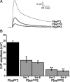
Figure 2.
Evoked release is reduced in phospholipid-binding mutants of both C2 domains. A, Representative traces recorded from larval muscle fiber 6. Each trace represents the mean of 30 consecutive sweeps from the same muscle fiber. B, Compared with P[sytWT], the mean EJP amplitude of all P[sytRQ] lines was significantly decreased (*, **p < 0.0001, 1-way ANOVA; P[sytWT], n = 16; P[sytA-RQ] line 7, n = 12; P[sytA-RQ] line 8, n = 14; P[sytB-RQ] line 3, n = 16; P[sytB-RQ] line 4, n = 13). Additionally, the evoked responses of the P[sytB-RQ] lines were significantly lower than those of the P[sytA-RQ] lines (*, **p < 0.01), but no differences were found between P[sytA-RQ] lines 7 and 8, or between P[sytB-RQ] lines 3 and 4 (p > 0.2).
Figure 3.
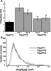
Figure 3.
mEJP frequency is increased in phospholipid-binding mutants of both C2 domains. A, Mean mEJP frequencies of each mutant and the transgenic control. P[sytWT], n = 55 fibers; P[sytA-RQ] line 7, n = 7 fibers; P[sytA-RQ] line 8, n = 17 fibers; P[sytB-RQ] line 3, n = 27 fibers; P[sytB-RQ] line 4, n = 12 fibers. Compared with P[sytWT], all genotypes had an increased mEJP frequency (p < 0.05). No significant difference was detected between P[sytA-RQ] and P[sytB-RQ] (p > 0.05), between P[sytA-RQ] lines 7 and 8 (p > 0.1), or between P[sytB-RQ] lines 3 and 4 (p > 0.1). *p < 0.0001, **p < 0.05, compared with P[sytWT]. B, Frequency distribution curves of mEJP amplitudes calculated from 1000 individual events per transgenic line in 0.2 mV bins. P[sytWT], P[sytA-RQ] line 8, and P[sytB-RQ] line 3 are shown.
Figure 4.
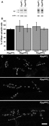
Figure 4.
Synaptotagmin expression is unaltered in phospholipid-binding mutants. A, Synaptotagmin is expressed at similar levels in each of the transgenic synaptotagmin lines. Representative Western blots of homogenized CNSs of third instars from the indicated lines were probed with an anti-synaptotagmin antibody. To confirm equal loading, they were also probed with an anti-actin antibody. B, Comparison of synaptotagmin/actin ratio normalized to the mean ratio of the transgenic control for P[sytWT] (n = 45), P[sytA-RQ] line 7 (n = 6), P[sytA-RQ] line 8 (n = 14), P[sytB-RQ] line 3 (n = 18), and P[sytB-RQ] line 4 (n = 10). An ANOVA showed no significant difference between any of the genotypes (p > 0.1). C, Synaptotagmin is localized to the larval neuromuscular junction in each of the transgenic synaptotagmin lines. Representative confocal Z-stack projections are shown for P[sytWT], P[sytA-RQ] line 8, and P[sytB-RQ] line 4. Scale bar, 10 μm.
Figure 5.
Figure 5.
Phospholipid-binding mutants decrease the apparent Ca2+ affinity but do not affect the Ca2+ cooperativity of release. A, EJPs were evoked in P[sytWT], P[sytA-RQ] line 8, and P[sytB-RQ] lines 3 and 4 by 0.05 Hz stimulation, and 10 sweeps were averaged for each fiber at each [Ca2+]. The responses from lines 3 and 4 of P[sytB-RQ] were not significantly different, so the results were pooled. For all genotypes at all [Ca2+], n = 10–18 muscle fibers, except for 1.5 mm Ca2+, at which n = 40–49 muscle fibers. The Hill equation was fit to the data. Error bars are SEM. B, The EJP amplitudes within the nonsaturating range of Ca2+ were plotted on a double-log plot, and a linear regression line was used to determine the slope (n) (P[sytWT]: n = 3.2, r = 0.999; P[sytA-RQ]: n = 3.0, r = 0.999; P[sytB-RQ]: n = 2.9, r = 0.933). C, The EJP amplitudes at each Ca2+ concentration were normalized to the maximum predicted by the Hill equation for each genotype and replotted to illustrate the shift in EC50. P[sytwt]: EC50 = 1.4 ± 0.1 mm; P[sytA-RQ]: EC50 = 2.0 ± 0.1 mm; P[sytB-RQ]: EC50 = 2.3 ± 0.2 mm. For all panels, black filled circles indicate P[sytWT], gray open squares indicate P[sytA-RQ], and gray open diamonds indicate P[sytB-RQ].
Figure 6.
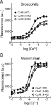
Figure 6.
Mutation of the conserved basic residue in either C2A or C2B decreases Ca2+-dependent interactions between anionic phospholipids and synaptotagmin in both Drosophila and mammals. Phospholipid binding for wild-type (black filled circles), C2A mutant (gray open squares; A-RQ in both Drosophila and mammals), and C2B mutant (gray open diamonds; B-RQ in Drosophila and B-KQ in mammals) C2AB domains are graphed versus Ca2+ concentration. A, Immobilized WT or A-RQ or B-RQ mutant versions of Drosophila C2AB were assayed for binding PS/PC liposomes. The A-RQ and the B-RQ mutations each decreased the apparent Ca2+ affinity of binding by ∼1.5-fold compared with WT without altering the Hill coefficient. The EC50 (mean ± SD in micromolar concentration) was 368 ± 35 for A-RQ, 344 ± 32 for B-RQ, and 238 ± 20 for WT. B, Immobilized WT, A-RQ, or B-KQ mutant versions of mammalian C2AB were assayed for binding PS/PC liposomes. The A-RQ and the B-KQ mutations each decreased the apparent Ca2+ affinity of binding by ∼1.5-fold compared with WT without altering the Hill coefficient. The EC50 (mean ± SD in micromolar concentration) was 154 ± 10 for A-RQ, 147 ± 7 for B-KQ, and 98 ± 6 for WT.
Figure 7.
Figure 7.
Model of the role played by the conserved basic residues in Ca2+-dependent interactions with the anionic presynaptic membrane. The crystal structure of the core complex [PDB file 1SFC, containing syntaxin (red), SNAP-25 (green), and VAMP (vesicle-associated membrane protein)/synaptobrevin (blue)], the nuclear magnetic resonance structures of the C2A (PDB file 1BYN) and C2B (PDB file 1K5W) domains of synaptotagmin (yellow), and Ca2+ (pink) are shown to scale using PyMOL Molecular Graphics System (DeLano Scientific). The membranes, the transmembrane domains, and the link between C2A and C2B were added in Adobe Photoshop. Left, A Ca2+-independent priming interaction between the C2B polylysine motif (yellow, space-filled residues) and SNAP-25 (green, space-filled residues) holds the C2A and C2B Ca2+ binding sites in close proximity to the presynaptic membrane. We diagrammed the interaction between the C2B polylysine motif and SNAP-25 within the SNARE complex (Zhang et al., 2002; Rickman et al., 2004, 2006; Dai et al., 2007), because mutation of the C2B polylysine motif impairs this interaction (Bai et al., 2004) and disrupts synaptotagmin's ability to increase the speed of SNARE-mediated liposome fusion in the absence of any PIP2 (Loewen et al., 2006). However, an interaction with PIP2, which is located specifically in the presynaptic membrane, could also serve this purpose (Bai et al., 2004; Araç et al., 2006). In the absence of Ca2+, the high concentration of negative charge in the Ca2+-binding pockets repulses the negatively charged presynaptic membrane, preventing synaptotagmin's conserved basic residues (blue, space-filled residues) from interacting with the membrane. Right, After Ca2+ entry, the negative charge of the Ca2+-binding pockets is neutralized by the bound Ca2+, which initiates the electrostatic switch: a strong attraction of the negatively charged, phospholipid head groups by the bound Ca2+ and the basic residues at the tips of Ca2+-binding pockets that draws the synaptic vesicle (SV) toward the presynaptic membrane (PM). Insertion of the hydrophobic residues (gray, stick residues) at the tips of the C2 domains into the core of the presynaptic membrane (Chapman and Davis, 1998) may help trigger fusion by promoting a local Ca2+-dependent buckling of the plasma membrane (Chapman and Davis, 1998; Martens et al., 2007). The Ca2+-induced increase in positive charge at the end of the C2B domain also likely increases the strength of the electrostatic interaction between the C2B polylysine motif and the SNARE complex, resulting in simultaneous binding of the SNARE complex and the presynaptic membrane (Davis et al., 1999; Bhalla et al., 2006; Loewen et al., 2006; Dai et al., 2007).

Similar articles

Cited by

References

    1. Araç D, Chen X, Khant HA, Ubach J, Ludtke SJ, Kikkawa M, Johnson AE, Chiu W, Südhof TC, Rizo J. Close membrane-membrane proximity induced by Ca(2+)-dependent multivalent binding of synaptotagmin-1 to phospholipids. Nat Struct Mol Biol. 2006;13:209–217. - PubMed
    1. Bai J, Wang P, Chapman ER. C2A activates a cryptic Ca(2+)-triggered membrane penetration activity within the C2B domain of synaptotagmin I. Proc Natl Acad Sci U S A. 2002;99:1665–1670. - PMC - PubMed
    1. Bai J, Tucker WC, Chapman ER. PIP2 increases the speed of response of synaptotagmin and steers its membrane-penetration activity toward the plasma membrane. Nat Struct Mol Biol. 2004;11:36–44. - PubMed
    1. Banker G, Goslin K, editors. Culturing nerve cells. Ed 2. Cambridge, MA: MIT; 1998.
    1. Bekkers JM, Stevens CF. Presynaptic mechanism for long-term potentiation in the hippocampus. Nature. 1990;346:724–729. - PubMed

Publication types

MeSH terms

LinkOut - more resources