. 2008 Jul 23;28(30):7687-98.
doi: 10.1523/JNEUROSCI.0143-07.2008.

Neuroinflammation and oxidation/nitration of alpha-synuclein linked to dopaminergic neurodegeneration

Affiliations

Neuroinflammation and oxidation/nitration of alpha-synuclein linked to dopaminergic neurodegeneration

Hui-Ming Gao et al. J Neurosci. .

Abstract

alpha-Synuclein (SYN) is the major component of Lewy bodies, the neuropathological hallmarks of Parkinson's disease (PD). Missense mutations and multiplications of the SYN gene cause autosomal dominant inherited PD. Thus, SYN is implicated in the pathogenesis of PD. However, the mechanism whereby SYN promotes neurodegeneration remains unclear. Familial PD with SYN gene mutations are rare because the majority of PD is sporadic and emerging evidence indicates that sporadic PD may result from genetic and environmental risk factors including neuroinflammation. Hence, we examined the relationship between SYN dysfunction and neuroinflammation in mediating dopaminergic neurodegeneration in mice and dopaminergic neuronal cultures derived from wild-type SYN and mutant A53T SYN transgenic mice in a murine SYN-null (SYNKO) background (M7KO and M83KO, respectively). Stereotaxic injection of an inflammagen, lipopolysaccharide, into substantia nigra of these SYN genetically engineered mice induced similar inflammatory reactions. In M7KO and M83KO, but not in SYNKO mice, the neuroinflammation was associated with dopaminergic neuronal death and the accumulation of insoluble aggregated SYN as cytoplasmic inclusions in nigral neurons. Nitrated/oxidized SYN was detected in these inclusions and abatement of microglia-derived nitric oxide and superoxide provided significant neuroprotection in neuron-glia cultures from M7KO mice. These data suggest that nitric oxide and superoxide released by activated microglia may be mediators that link inflammation and abnormal SYN in mechanisms of PD neurodegeneration. This study advances understanding of the role of neuroinflammation and abnormal SYN in the pathogenesis of PD and opens new avenues for the discovery of more effective therapies for PD.


Figures
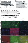
Figure 1.
Figure 1.
Expression of human SYN in Tg mice. A, Genotype analyses of tail DNA from homozygous mice by PCR amplification and slot blotting (SB). B, Western blot analysis of SYN expression in 3-month-old mice. The brain tissues were lysed in 2% SDS and 50 mm Tris-HCl, size fractionated by 12% SDS-PAGE gels, probed by Western blot for SNL1 (specific for mouse and human SYN), LB509 (specific for human SYN), or actin (as a loading control), and imaged by chemiluminescence. C, The cell-type specificity of SYN expression. The protein extracts from highly enriched neurons (N), microglia (M), or astroglia (A) were analyzed by Western blotting using anti-SYN antibodies. SYN was only expressed in neurons in both nTg and Tg mice. The level of SYN protein was undetectable in either microglia or astroglia even when lager amount of protein was loaded on the SDS-PAGE gel. D, Cellular localization of SYN. Immunofluorescence using antibodies against SYN and/or synaptophysin, a presynaptic protein, indicated that SYN was expressed predominantly at presynaptic terminals of cultured midbrain neurons in both nTg and SYN Tg mice. mSYN, Mouse SYN; hSYN, human SYN; NEO, neomycin.
Figure 2.
Figure 2.
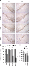
Differential vulnerability of nigral DA neurons to LPS-mediated neurotoxicity. A, Mouse brain sections were immunolabeled with antibody against TH and counterstained with Nissl. Representative images indicate the SN lesion in the LPS-injected side in nTg, M7KO, and M83KO mice 4 weeks after LPS injection, whereas in SYNKO mice, no significant damage of DA neurons was observed in the LPS-injected side compared with the NS-injected side. The loss of nigral DA neurons was most prominent in M83KO mice. B, Comparison of the number of nigral TH-IR neurons between the LPS-injected and the NS-injected side indicated that no significant loss of TH-IR neurons was detected in SYNKO mice, whereas significant loss of DA neurons was found in LPS-injected SN in nTg and SYN Tg mice 4 weeks after the injection. C, Quantification of NeuN-IR neurons in the LPS-injected SN revealed a significant decrease in M83KO mice but not in M7KO mice. Results are expressed as a percentage of the NS-injected side of the corresponding genotype. Error bars indicate mean ± SEM. *p < 0.01, compared with the corresponding NS-injected control side; #p < 0.01, compared with the LPS-injected side of M7KO mice.
Figure 3.
Figure 3.
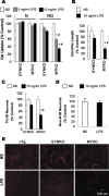
Overexpression of human SYN in neurons renders DA neurons more sensitive to inflammation. Midbrain neuron-enriched (N) or neuron–glia cultures (NG) from SYNKO or M7KO mice were treated for 7 d with NS or 5–10 ng/ml LPS. DA neurons were labeled with anti-TH antibody. A–C, Dopaminergic neurodegeneration was evaluated with [3H]DA uptake assay (A), measurement of dendrite length of individual TH-IR neurons (B), or quantification of TH-IR neurons in NG cultures (C). D, Quantification of the number of NeuN-IR neurons in midbrain NG cultures from M7KO mice 7 d after the treatment with 10 ng/ml LPS. The results are expressed as percentage of the control cultures and are the mean ± SEM of three to four experiments performed in triplicate. *p < 0.01, compared with the corresponding NS-treated NG cultures; #p < 0.01, compared with the corresponding LPS-treated NG cultures from SYNKO mice. E, Midbrain NG cultures were treated for 7 d with NS or LPS. Cultures then were immunostained for TH.
Figure 4.
Figure 4.
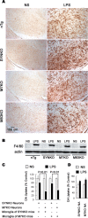
The differential degeneration of DA neurons is not attributable to different inflammatory reactions. A, LPS-induced activation of microglia in the mouse SN. One week after the injection, brain sections were immunostained with an antibody against F4/80 to determine the activation status of the microglia. In the NS-injected SN, microglia exhibited the typical ramified morphology of resting microglia. In the LPS-injected SN, microglia were highly activated with the characteristics of larger size, irregular shape, and dramatically increased expression of the F4/80 antigen. Note that the activation status of microglia appeared similar in all genotypes. B, Western blot analysis showed strikingly increased expression of F4/80 in the mouse midbrain 1 week after LPS injection. The levels of F4/80 expression were not different among different mouse genotypes. C, Midbrain neuron-enriched cultures from SYNKO or M7KO mice were supplemented with 5 × 10⁴ microglia per well prepared from either SYNKO or M7KO mice. After 24 h, the cultures were treated with NS or 10 ng/ml LPS, and [3H]DA uptake was determined 7 d after the treatment. p < 0.01 was considered statistically significant. Note that in these reconstituted cultures, the presence of human SYN in neurons rendered DA neurons more sensitive to inflammation, regardless of the origin of supplemented microglia. D, The neuron–astroglia cocultures (NA) were treated with NS or LPS, and [3H]DA uptake was determined 7 d after the treatment. The activation of astroglia after LPS treatment provided similar neuroprotection in cultures from SYNKO and M7KO mice. Error bars indicate SEM.
Figure 5.
Figure 5.
Microglia-derived superoxide and NO are major mediators of dopaminergic neurodegeneration. A–D, Neuron–glia cultures (A, D) and microglia-enriched cultures (B, C) were treated with NS or LPS. At the indicated time point, the production of TNFα (A), superoxide (B), and nitrite (an indicator of NO production) (C, D) was measured. The levels of these factors were dramatically increased with LPS stimulation, but they were not significantly different between two genotypes. E, In vivo iNOS expression was similarly increased 1 week after LPS injection in mouse midbrains. F, Abatement of NO and superoxide mitigated LPS-induced degeneration of DA neurons. Neuron–glia cultures were pretreated for 30 min with the iNOS inhibitor 7-nitroindazole or NADPH oxidase inhibitor apocynin followed with the treatment with NS or 10 ng/ml LPS for 7 d. Cultures were then assayed for DA uptake. Both 7-nitroindazole and apocynin provided significant neuroprotection in the cultures from M7KO mice. G, NMDA glutamate receptor antagonist MK801 attenuated LPS neurotoxicity in neuron–glia cultures prepared from M7KO mice. The cultures were pretreated for 30 min with 10 μm MK801 and the DA uptake assay was performed 7 d after the treatment with NS or 10 ng/ml LPS. The results are the mean ± SEM of two to three experiments performed in triplicate. *p < 0.01 compared with the corresponding LPS-treated cultures.
Figure 6.
Figure 6.
Accumulation of insoluble and aggregated SYN in mouse midbrain after LPS injection. A, Mouse brain sections were double labeled with anti-TH (green) and SYN211 (specific to human SYN, red) antibodies. SYN211 staining revealed that some of TH-expressing cells in the LPS-injected SN contained SYN aggregates. The inset and its magnified photomicrograph (bottom) display SYN aggregates in TH-expressing neurons in the LPS-injected SN. B, In neuron–glia cultures, SYN appeared mainly in perinuclear locations and formed aggregation 7 d after the LPS treatment. SYN505, antibody raised to oxidized human SYN, positively stained these aggregates. C, D, Midbrain tissues were sequentially extracted and size fractionated by 12% SDS-PAGE gels, followed by Western blot analysis using antibody SNL1 or LB509. Insoluble SYN was detected only in extracts of LPS-injected midbrains, but not in NS-injected midbrains. E, HST-insoluble fraction (dissolved in RIPA buffer) was size fractionated on a nondenaturing 12% polyacrylamide gel and probed by Western blotting for SNL1. The aggregated SYN was seen in the stacking gel in LPS-injected midbrain extracts, but not in NS-injected midbrain extracts. The arrow indicates the resolving and stacking gel interface. N, NS; L, LPS; DAPI, 4′,6′-diamidino-2-phenylindole dihydrochloride.
Figure 7.
Figure 7.
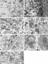
Neuronal degeneration associated with SYN pathology in the LPS-injected SN of M7KO mice. A, C, Unaffected neuron reveals normal morphology of mitochondria and other organelles. B, D, Myelin disruption and axonal degeneration in the LPS-injected nigral neurons. Myelin is fragmented and disorganized, and the axons within the myelin sheaths are severely atrophic (arrowheads). D, Interestingly, there is an abnormal membrane structure that appears in the nucleus of a damaged neuron (arrow). E–H, Anti-SYN antibody SYN211 staining associated with membrane structures showed damaged mitochondria (arrowheads) in affected neurons. SYN-positive silver grains also appeared to be associated with the lysosomes (arrow), Golgi apparatus (F), and multilamellar bodies (G). G, Magnified photomicrograph from boxed area (defined by solid black rectangles) in H shows the specifically labeled silver grains in the multilamellar bodies. Scale bars, 500 nm.
Figure 8.
Figure 8.
Pathogenic modifications of SYN in cytoplasmic inclusions. A, D, Brain sections were first stained with an anti-NeuN antibody using DAB as a chromophore and nickel sulfate as an intensifying agent (dark blue) followed by (A) nSYN14 antibody (specific for nitrated human SYN) or (D) pSER129 SYN antibody (specific for Ser129-phosphorylated SYN) using DAB as a chromophore (brown). nSYN14-positive cytoplasmic inclusions were observed in neurons in LPS-injected SN, but not in NS-injected SN in M7KO and M83KO mice. B, Western blot analyses using nSYN14 shows that nitrated SYN appeared in LPS-injected (L) midbrain extracts from both M7KO and M83KO mice, but not in NS-injected (N) midbrain samples. C, Blockade of NMDA glutamate receptors using MK801 (M) significantly reduced the nitration of SYN. Neuron–glia cultures prepared from M7KO mice were pretreated for 30 min with MK801 followed with the treatment with NS or 10 ng/ml LPS for 7 d. Cultures were then extracted with RIPA buffer and Western blot analysis using nSYN14 was used to detect the nitration of SYN. D, Phosphorylated SYN at Ser129 appeared in the neuronal inclusions in the LPS-injected SN of M7KO mice. E, Midbrain tissues were sequentially extracted 4 weeks after LPS/NS injection, and analyzed by Western blotting using LB509 or an antibody specific for Ser129-phosphorylated (pSER129) SYN. Ser129-phosphorylated SYN appeared in HST-insoluble fractions in LPS-injected midbrains, but not in NS-injected midbrains. In sequential extracts of midbrain from a PD patient, Ser129-phosphorylated SYN only could be detected in insoluble fractions (SDS fractions). C, Control; M+L, MK801 plus LPS.

Similar articles

Cited by

References

    1. Abeliovich A, Schmitz Y, Fariñas I, Choi-Lundberg D, Ho WH, Castillo PE, Shinsky N, Verdugo JM, Armanini M, Ryan A, Hynes M, Phillips H, Sulzer D, Rosenthal A. Mice lacking alpha-synuclein display functional deficits in the nigrostriatal dopamine system. Neuron. 2000;25:239–252. - PubMed
    1. Arawaka S, Wada M, Goto S, Karube H, Sakamoto M, Ren CH, Koyama S, Nagasawa H, Kimura H, Kawanami T, Kurita K, Tajima K, Daimon M, Baba M, Kido T, Saino S, Goto K, Asao H, Kitanaka C, Takashita E, et al. The role of G-protein-coupled receptor kinase 5 in pathogenesis of sporadic Parkinson's disease. J Neurosci. 2006;26:9227–9238. - PMC - PubMed
    1. Bal-Price A, Brown GC. Inflammatory neurodegeneration mediated by nitric oxide from activated glia-inhibiting neuronal respiration, causing glutamate release and excitotoxicity. J Neurosci. 2001;21:6480–6491. - PMC - PubMed
    1. Barger SW, Goodwin ME, Porter MM, Beggs ML. Glutamate release from activated microglia requires the oxidative burst and lipid peroxidation. J Neurochem. 2007;101:1205–1213. - PMC - PubMed
    1. Block ML, Wu X, Pei Z, Li G, Wang T, Qin L, Wilson B, Yang J, Hong JS, Veronesi B. Nanometer size diesel exhaust particles are selectively toxic to dopaminergic neurons: the role of microglia, phagocytosis, and NADPH oxidase. FASEB J. 2004;18:1618–1620. - PubMed

Publication types

MeSH terms