. 2008 Sep 26;283(39):26759-70.
doi: 10.1074/jbc.M802160200. Epub 2008 Jul 23.

A novel mechanism by which thiazolidinediones facilitate the proteasomal degradation of cyclin D1 in cancer cells

Affiliations

A novel mechanism by which thiazolidinediones facilitate the proteasomal degradation of cyclin D1 in cancer cells

Shuo Wei et al. J Biol Chem. .

Abstract

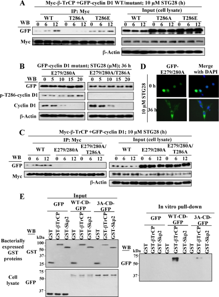
This study identifies a novel mechanism by which thiazolidinediones mediate cyclin D1 repression in prostate cancer cells. Based on the finding that the thiazolidinedione family of peroxisome proliferator-activated receptor gamma (PPARgamma) agonists mediated PPARgamma-independent cyclin D1 degradation, we developed a novel PPARgamma-inactive troglitazone derivative, STG28, with high potency in cyclin D1 ablation. STG28-mediated cyclin D1 degradation was preceded by Thr-286 phosphorylation and nuclear export, which however, were independent of glycogen synthase kinase 3beta. Mutational analysis further confirmed the pivotal role of Thr-286 phosphorylation in STG28-induced nuclear export and proteolysis. Of several kinases examined, inhibition of IkappaB kinase alpha blocked STG28-mediated cytoplasmic sequestration and degradation of cyclin D1. Pulldown of ectopically expressed Cul1, the scaffold protein of the Skp-Cullin-F-box E3 ligase, in STG28-treated cells revealed an increased association of cyclin D1 with beta-TrCP, whereas no specific binding was noted with other F-box proteins examined, including Skp2, Fbw7, Fbx4, and Fbxw8. This finding represents the first evidence that cyclin D1 is targeted by beta-TrCP. Moreover, beta-TrCP expression was up-regulated in response to STG28, and ectopic expression and small interfering RNA-mediated knock-down of beta-TrCP enhanced and protected against STG28-facilitated cyclin D1 degradation, respectively. Because cyclin D1 lacks the DSG destruction motif, mutational and modeling analyses indicate that cyclin D1 was targeted by beta-TrCP through an unconventional recognition site, (279)EEVDLACpT(286), reminiscent to that of Wee1. Moreover, we obtained evidence that this beta-TrCP-dependent degradation takes part in controlling cyclin D1 turnover when cancer cells undergo glucose starvation, which endows physiological relevance to this novel mechanism.


Figures

FIGURE 1.
FIGURE 1.
STG28 facilitates cyclin D1 phosphorylation, nuclear export, and proteasomal degradation. A, chemical structure of troglitazone (TG) and STG28 and Western blot analyses of the dose-dependent effect of these two thiazolidinediones on suppressing cyclin D1 expression in LNCaP cells in 10% FBS-supplemented RPMI 1640 medium for 72 h. B, protective effect of proteasomal inhibitor, MG132, on STG28-mediated cyclin D1 degradation (left panel). LNCaP cells were exposed to 10 μm STG28 alone or in combination with 10 μm MG132 in 10% FBS-supplemented medium for the indicated time intervals followed by immunoblotting with anti-cyclin D1 antibodies. Right panel, STG28 does not affect cyclin D1 mRNA levels. RT, reverse transcription. C, STG28 shortens cyclin D1 protein half-life. Cells were pretreated with either DMSO or 10 μm STG28 for 6 h followed by exposure to 100 μg/ml cycloheximide for the indicated time intervals followed by immunoblotting with anti-cyclin D1 antibodies. D, time-dependent effect of 10 μm STG28 on cyclin D1 phosphorylation at the Thr-286 residue with normalized cyclin D1 levels. E, immunocytochemical evidence of STG28-induced nuclear export of cyclin D1. Left panel, intracellular distribution of endogenous cyclin D1 after treating LNCaP cells with 10 μm STG28 for the indicated time intervals. Right panel, time-dependent cytoplasmic sequestration of ectopically expressed GFP-cyclin D1 in response to STG28 treatment. DAPI, 4,6-diamidino-2-phenylindole.
FIGURE 2.
FIGURE 2.
STG28-mediated cyclin D1 degradation requires nuclear export and is GSK3β- and cell cycle-independent. A, time-dependent effect of STG28 on modulating nuclear versus cytosolic cyclin D1 in LNCaP cells. Cells were treated with 10 μm STG28 in 10% FBS-containing medium for the indicated time intervals. Cell lysates were fractionated into cytoplasmic and nuclear fractions followed by immunoblotting with anti-cyclin D1 antibodies with β-actin and nucleolin as internal references, respectively. B, leptomycin B (LMB), an inhibitor of CRM1-dependent nuclear export, provides a dose-dependent protection against STG28-mediated cyclin D1 degradation after 36 h of incubation. C, STG28-mediated cyclin D1 degradation is GSK3β-independent. D, flow cytometry analysis of the time-dependent effect of 10 μm STG28 on altering cell cycle distribution in LNCaP cells in 10% FBS-containing RPMI 1640 medium.
FIGURE 3.
FIGURE 3.
Evidence that the Thr-286 residue is integral to STG28-facilitated nuclear export and proteolysis of cyclin D1. A, upper panel, schematic representation of the structure of the GFP-cyclin D1 fusion protein. Lower panel, immunoblotting (IB) of GFP-cyclin D1 and various GFP-cyclin D1 mutants ectopically expressed in LNCaP cells. B, left panel, immunocytochemical analysis of the subcellular distribution of various GFP-cyclin D1 mutants in DMSO- and STG28-treated LNCaP cells. GFP-cyclin D1 mutants were ectopically expressed in LNCaP cells, and their subcellular localization was visualized by fluorescence microscopy after 36 h of treatment with DMSO or 10 μm STG28. Right panel, percentage nuclear distribution of GFP-tagged wild-type (WT) versus mutant cyclin D1 in STG28-treated LNCaP cells relative to that of DMSO-treated cells. Three random fields were inspected, and 10-20 transfected cells were counted in each field. Columns, mean (n = 3); bar, S.D. C, dose-dependent effect of STG28 on the degradation of the various GFP-cyclin D1 mutants in LNCaP cells. Cells ectopically expressing GFP-cyclin D1 mutants were treated with STG28 at the indicated concentrations for 48 h and immunoblotted for GFP and cyclin D1. Endogenous cyclin D1 degradation was used as an internal control for STG28 activity. DAPI, 4,6-diamidino-2-phenylindole.
FIGURE 4.
FIGURE 4.
Evidence that IKKα plays a pivotal role in STG28-mediated cyclin D1 nuclear export and degradation. A, left panel, time-dependent effect of 10 μm STG28 on the phosphorylation status of GSK3β, IKKα, ERKs, and p38 in LNCaP cells in 10% FBS-containing medium for 36 h. Right panel, effects of specific kinase inhibitors (PD98059, ERKs; SB203580, p38; Bay11-7082, IKKα) on STG28-mediated cyclin D1 degradation. LNCaP cells were exposed to STG28 and/or individual kinase inhibitors at the indicated concentrations for 36 h. B, siRNA-mediated knockdown of IKKα rescues STG28-induced cyclin D1 repression. C, IKKα inhibition by the ectopic expression of a dominant negative IKKα mutant (IKK2M) protected cyclin D1 from STG28-induced degradation. Percentages represent the relative expression levels of cyclin D1 compared with that at the 0-h time point and normalized to β-actin. D, immunocytochemical evidence that ectopic expression of IKK2M hindered STG28-mediated GFP-cyclin D1 nuclear export. DAPI, 4,6-diamidino-2-phenylindole.
FIGURE 5.
FIGURE 5.
Role of SCFβ-TrCP in STG28-facilitated cyclin D1 proteolysis. A, cyclin D1 co-immunoprecipitated (IP) with the SCF complex scaffold protein Cul1 and the F-box protein β-TrCP in STG28-treated LNCaP cells. LNCaP cells were transiently transfected with Cul1-FLAG plasmids and treated with 10 μm STG28 for 8 or 20 h followed by co-treatment with MG132 for an additional 4 h. Immunoprecipitation with anti-cyclin D-agarose conjugates and immunoblotting for cyclin D1, Cul1 (FLAG), and F-box proteins β-TrCP, Skp2, Fbw7, Fbx4, and Fbxw8 were performed as described in the “Experimental Procedures.” B, immunocytochemical evidence that STG28 promoted the cytoplasmic co-localization of cyclin D1 and β-TrCP in LNCaP cells. LNCaP cells were treated with DMSO or 10 μm STG28 for 36 h, and the subcellular localization of cyclin D1 and β-TrCP was visualized by fluorescence microscopy. C, pulldown analysis of the selective binding of cyclin D1 to β-TrCP. LNCaP cells were transiently transfected with Myc-tagged β-TrCP, Skp2, or Fbw7 and treated with MG132 for 4 h followed by anti-Myc-agarose. The complex was immunoblotted (WB) with antibodies against cyclin D1, β-catenin, and Myc as described under “Experimental Procedures.” D, Western blot analysis of the dose-dependent effects of STG28 on modulating the expression levels of cyclin D1 and the F-box proteinsβ-TrCP, Skp2, Fbw7, Fbx4, and Fbxw8 (left panel) as well as known β-TrCP substrates, including β-catenin, Wee1, IκBα, and Cdc25A (right panel) in LNCaP cells. Percentages represent the relative expression levels of cyclin D1 and F-box proteins compared with that in the DMSO group and normalized to β-actin.
FIGURE 6.
FIGURE 6.
Evidence that β-TrCP is involved in STG28-induced cyclin D1 degradation. A, left panel, ectopic β-TrCP expression facilitated cyclin D1 degradation in a manner similar to STG28. LNCaP cells were nucleofected with increased concentrations of Myc-tagged β-TrCP or Skp2 for 36 h. Cell lysates were collected and subjected to immunoblotting with cyclin D1 and Myc antibodies. Right panel, overexpression of β-TrCP enhanced STG28-mediated cyclin D1 proteolysis. LNCaP cells were transiently transfected with pCMV or β-TrCP-Myc plasmids and treated 10 μm STG28 for the indicated time intervals. Cell lysates were immunoblotted with antibodies against cyclin D1 and Myc. Percentages represent the relative expression levels of cyclin D1 compared with that at the 0-h time point and normalized to β-actin. B and C, siRNA-mediated knockdown of β-TrCP, but not Fbxw8, rescued STG28-induced cyclin D1 degradation. LNCaP cells were transiently transfected with scrambled (Scrb), β-TrCP, or Fbxw8 siRNA and treated with 10 μm STG28 for 72 h. Cell lysates were immunoblotted with antibodies against cyclin D1, β-TrCP, and Fbxw8 (upper panels). These immunoblots are representative of three independent experiments. Lower panels, relative percentages of cyclin D1 expression levels in LNCaP cells with different treatments to those of untreated cells. Columns, mean (n = 3); bar, S.D. D, overexpression of β-TrCP enhanced STG28-mediated cyclin D1 ubiquitination. Cells were co-nucleofected with 5 μg of HA-ubiquitin with vector control pCMV or β-TrCP-Myc and incubated in 10% FBS-containing medium for 24 h followed by STG28 treatment for the indicated time intervals. Equal amounts of cell lysates were probed with anti-cyclin D1 and anti-Myc antibodies (Input) or immunoprecipitated (IP) with anti-HA or anti-FLAG affinity matrix followed by Western blot (WB) analysis with anti-cyclin D1 antibodies.
FIGURE 7.
FIGURE 7.
Mutational analyses of the pivotal role of the triad Thr-286, Glu-279, and Glu-280 in theβ-TrCP recognition and STG28-mediated degradation of cyclin D1. A, involvement of Thr-286 in cyclin D1 recognition by β-TrCP. LNCaP cells ectopically expressing both Myc-tagged β-TrCP (Myc-β-TrCP) and GFP-tagged cyclin D1 variant (wild-type (WT), T286A, or T286E) were treated with 10 μm STG28 for 2 or 8 h followed by co-treatment with MG132 for 4 h. Immunoprecipitation (IP) with anti-Myc-agarose conjugates and immunoblotting (WB) for GFP and Myc were performed as described under “Experimental Procedures.” B, GFP-cyclin D1 E279A/E280A and E279A/E280A/T286A mutants were resistant to STG28-induced cyclin D1 proteolysis. LNCaP cells expressing E279A/E280A or E279A/E280A/T286A mutants were treated with different doses of STG28 for 36 h, and cell lysates were immunoblotted with antibodies against GFP, p-Thr-286-cyclin D1, and cyclin D1. C, GFP-cyclin D1 E279A/E280A and E279A/E280A/T286A mutants were incapable of binding to β-TrCP. LNCaP cells ectopically expressing GFP-cyclin D1 wild type or E279A/E280A or E279A/E280A/T286A mutants were treated with 10 μm STG28 for 2 or 8 h followed by a 4-h cotreatment with MG132. Immunoprecipitation with anti-Myc-agarose conjugates and immunoblotting for GFP and Myc were performed as described under “Experimental Procedures.” D, GFP-cyclin D1 E279A/E280A mutant was localized to the cytoplasm in STG28-treated cells. LNCaP cells expressing the GFP-cyclin D1 E279A/E280A mutant were treated with 10 μm STG28 for 36 h, and the subcellular localization of GFP-cyclin D1 was visualized by fluorescence microscopy. E, in vitro pulldown of GFP-cyclin D1 by bacterially expressed GST-β-TrCP. LNCaP cells were transfected with GFP, wild-type cyclin D1-GFP (WT-CD-GFP), or E279A/E280A/T286A cyclin D1-GFP (3A-CD-GFP) for 24 h, and equal amounts of cell lysates were incubated with recombinant GST, GST-β-TrCP, or GST-Skp2 immobilized onto glutathione beads. The resulting complexes were washed, centrifuged, and subjected to Western blot analysis with GFP antibodies (right panel). One-tenth of cell lysates were collected as input and probed with GFP antibodies, and recombinant GST-fusion proteins (GST, GST-β-TrCP, and GST-Skp2) were purity- and protein integrity-confirmed with GST antibody (left panel).
FIGURE 8.
FIGURE 8.
Molecular modeling analysis of β-TrCP WD40 domain and β-TrCP binding motif of cyclin D1, 279EEVDLACpT286. Interface structures of the doubly phosphorylated β-catenin peptide (left panel) and the singly phosphorylated cyclin D1 peptide (right panel) bound to the β-TrCP1 WD40 domain. The β-TrCP1 WD40 domain is shown as solid ribbons, with its involved side chains in stick form, and the peptides of β-catenin and cyclin D1 are represented as green and orange tubes, respectively, with their identified side chains shown in light green and yellow stick forms, respectively.
FIGURE 9.
FIGURE 9.
STG28 mimics the effect of glucose starvation on cyclin D1 degradation. A, dose-dependent effects of STG28 and time-dependent effect of glucose starvation on modulating the expression of cyclin D1, β-TrCP, and its known substrates, including β-catenin, IκBα, and Wee1. LNCaP cells were exposed to different concentrations of STG28 for 72 h or incubated in glucose-deprived, 10% FBS-supplemental RPMI 1640 medium for the indicated time intervals, and immunoblotting was performed as described under “Experimental Procedure.” B, glucose starvation promoted the association of cyclin D1 with β-TrCP. Cells were incubated in glucose-deprived medium for different time intervals followed by immunoprecipitation (IP) with anti-cyclin D1-agarose, and immunoblotting for cyclin D1,β-TrCP, Skp2, Fbw7, Fbx4, and Fbxw8 were performed.

Similar articles

Cited by

References

    1. Diehl, J. A. (2002) Cancer Biol. Ther. 1226 -231 - PubMed
    1. Weinstein, I. B. (2002) Science 29763 -64 - PubMed
    1. Stacey, D. W. (2003) Curr. Opin. Cell Biol. 15158 -163 - PubMed
    1. Chung, D. C. (2004) Ann. N. Y. Acad. Sci. 1014209 -217 - PubMed
    1. Fu, M., Wang, C., Li, Z., Sakamaki, T., and Pestell, R. G. (2004) Endocrinology 1455439 -5447 - PubMed

Publication types

MeSH terms

Substances