. 2008 Sep 19;283(38):25928-35.
doi: 10.1074/jbc.M801326200. Epub 2008 Jul 23.

Activation of Polo-like kinase 3 by hypoxic stresses

Affiliations

Activation of Polo-like kinase 3 by hypoxic stresses

Ling Wang et al. J Biol Chem. .

Abstract

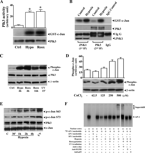
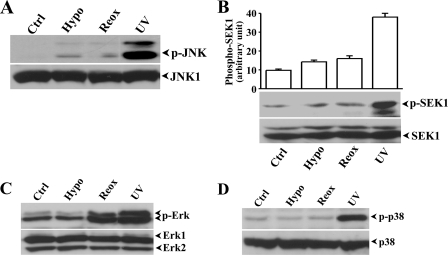
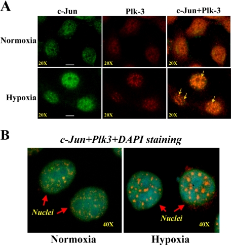
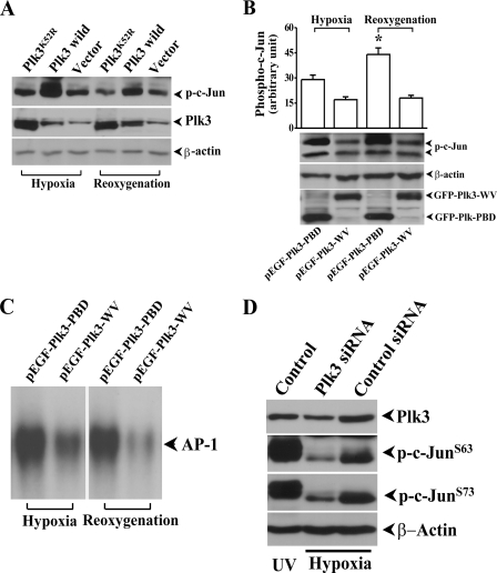
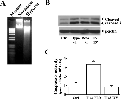
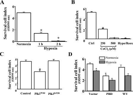
Hypoxia/reoxygenation stress induces the activation of specific signaling proteins and activator protein 1 (AP-1) to regulate cell cycle regression and apoptosis. In the present study, we report that hypoxia/reoxygenation stress activates AP-1 by increasing c-Jun phosphorylation and DNA binding activity through activation of Polo-like-kinase 3 (Plk3) resulting in apoptosis. The specific effect of hypoxia/reoxygenation stress on Plk3 activation resulting in c-Jun phosphorylation was the opposite of UV irradiation-induced responses that are meanly independent on activation of the stress-induced JNK signaling pathway in human corneal epithelial (HCE) cells. The effect of hypoxia/reoxygenation stress-induced Plk3 activation on increased c-Jun phosphorylation and apoptosis was also mimicked by exposure of cells to CoCl(2). Hypoxia/reoxygenation activated Plk3 in HCE cells to directly phosphorylate c-Jun proteins at phosphorylation sites Ser-63 and Ser-73, and to increase DNA binding activity of c-Jun, detected by EMSA. Further evidence demonstrated that Plk3 and phospho-c-Jun were immunocolocalized in the nuclear compartment of hypoxia/reoxygenation stress-induced cells. Increased Plk3 activity by overexpression of wild-type and dominantly positive Plk3 enhanced the effect of hypoxia/reoxygenation on c-Jun phosphorylation and cell death. In contrast, knocking-down Plk3 mRNA suppressed hypoxia-induced c-Jun phosphorylation. Our results provide a new mechanism indicating that hypoxia/reoxygenation induces Plk3 activation instead of the JNK effect to directly phosphorylate and activate c-Jun, subsequently contributing to apoptosis in HCE cells.


Figures

FIGURE 1.
FIGURE 1.
Hypoxia/reoxygenation-induced activation of Plk3 and c-Jun phosphorylation in HCE Cells. A, effect of hypoxia (Hypo) and reoxygenation (Reox) on Plk3 activity. Immunocomplex kinase assays were performed to determine Plk3 kinase activity, and hypophosphorylated GST-c-Jun fusion protein was used as a substrate. B, effect of hypoxia-activated Plk3 on c-Jun phosphorylation after removing JNK. JNK protein was removed from cell lysates by immunoprecipitation, and Plk3 activity was measured by immunocomplex kinase assay. C, hypoxia/reoxygenation-induced Plk3 activation and c-Jun phosphorylation. Western blots were used to detect Plk3 expression and phosphorylated c-Jun using antibody specific to phospho-c-Jun. D, dose-dependent response of CoCl2-induced phosphorylation of c-Jun. E, time course of hypoxia-induced site-specific phosphorylation of c-Jun at Ser-63 and Ser-73. F, effect of hypoxia and reoxygenation on AP-1 DNA binding activity detected by EMSA and supershift. UV irradiation-induced phosphorylation of c-Jun and AP-1 activation were detected as positive controls.
FIGURE 2.
FIGURE 2.
Hypoxia/reoxygenation-induced activation of MAP kinases. Effect of hypoxia (Hypo) and reoxygenation (Reox) on: A, JNK phosphorylation; B, SEK phosphorylation; C, Erk phosphorylation; and D, p38 phosphorylation. UV irradiation-induced MAP kinase activation served as positive controls. Activation of SEK and MAP kinases were determined by detecting hypoxia-induced changes in phosphorylation levels of these proteins with specific anti-phospho-antibodies. Statistical significance was indicated by symbols of * and ** for p < 0.05 and p < 0.01, respectively.
FIGURE 3.
FIGURE 3.
Immunocolocalization of Plk3 and c-Jun. A, co-localization of Plk3 and c-Jun in hypoxia-induced cells. B, co-localization of Plk3 and c-Jun in nuclei. Cellular Plk3 and c-Jun proteins in control and hypoxia-induced HCE cell were stained using monoclonal antibodies specific to Plk3 and c-Jun, respectively. Cell nuclei were stained by DAPI.
FIGURE 4.
FIGURE 4.
Effect of Plk3 mutants on c-Jun phosphorylation and AP-1 activation. A, effect of overexpressing wild-type Plk3 and kinase-defective Plk3 mutant (Plk3K52R) on c-Jun phosphorylation. B, effect of overexpressing constitutively active Plk3 (Plk3-PBD) and defective Plk3 (Plk3-WV) on c-Jun phosphorylation. C, effect of overexpressing constitutively active Plk3 (Plk3-PBD) and defective Plk3 (Plk3-WV) on AP-1 activation detected by EMSA. Control experiments were performed by transfecting HCE cells with GFP-tagged Plk3 and the vector only. D, effect of knocking-down Plk3 using siRNA on hypoxia-induced c-Jun phosphorylation. UV irradiation-induced c-Jun phosphorylation served as positive controls. The symbol * indicates a significant difference (p < 0.05, n = 3).
FIGURE 5.
FIGURE 5.
Effect of Plk3 on hypoxia-induced apoptosis. A, detection of hypoxia-induced DNA fragmentation. B, activation of caspase 3 induced by hypoxia and reoxygenation. UV irradiation-induced caspase 3 activation served as the positive control. C, effect of overexpressing constitutively active Plk3 (Plk3-PBD) and defective Plk3 (Plk3-WV) on caspase 3 activation. The symbol * indicates a significant difference (p < 0.01, n = 4).
FIGURE 6.
FIGURE 6.
Effect of Plk3 on hypoxia/reoxygenation-induced changes in survival cell index detected by MTT assay. A, hypoxia-induced decrease in survival cell index. B, CoCl2 induced a decrease in survival cell index. C, effect of overexpressing wild-type Plk3 and kinase-defective Plk3 mutant (Plk3K52R) on survival cell index. D, effect of overexpressing constitutively active Plk3 (Plk3-PBD) and defective Plk3 (Plk3-WV) on hypoxia-induced survival cell index. The symbol * indicates a significant difference (p < 0.05, n = 4).

Similar articles

Cited by

References

    1. Singh, N., Amin, S., Richter, E., Rashid, S., Scoglietti, V., Jani, P. D., Wang, J., Kaur, R., Ambati, J., Dong, Z., and Ambati, B. K. (2005) Investig. Ophthalmol. Vis. Sci. 46 1647–1652 - PubMed
    1. Singh, N., Jani, P. D., Suthar, T., Amin, S., and Ambati, B. K. (2006) Investig. Ophthalmol. Vis. Sci. 47 4787–4793 - PubMed
    1. Cursiefen, C., Maruyama, K., Jackson, D. G., Streilein, J. W., and Kruse, F. E. (2006) Cornea 25 443–447 - PubMed
    1. Mwaikambo, B. R., Sennlaub, F., Ong, H., Chemtob, S., and Hardy, P. (2006) Invest Ophthalmol. Vis. Sci. 47 4356–4364 - PubMed
    1. Zhang, M. C., Wang, Y., and Yang, Y. (2006) Ocul. Immunol. Inflamm. 14 359–365 - PubMed

Publication types