

doi: 10.1074/jbc.M804272200.
Epub 2008 Jul 23.
Collagen fibril formation. A new target to limit fibrosis
Affiliations
- PMID: 18650436
- PMCID: PMC2533774
- DOI: 10.1074/jbc.M804272200
Item in Clipboard
Collagen fibril formation. A new target to limit fibrosis
J Biol Chem.
.
Abstract
We present a concept for reducing formation of fibrotic deposits by inhibiting self-assembly of collagen molecules into fibrils, a main component of fibrotic lesions. Employing monoclonal antibodies that bind to the telopeptide region of a collagen molecule, we found that blocking telopeptide-mediated collagen/collagen interactions reduces the amount of collagen fibrils accumulated in vitro and in keloid-like organotypic constructs. We conclude that inhibiting extracellular steps of the fibrotic process provides a novel approach to limit fibrosis in a number of tissues and organs.
Figures
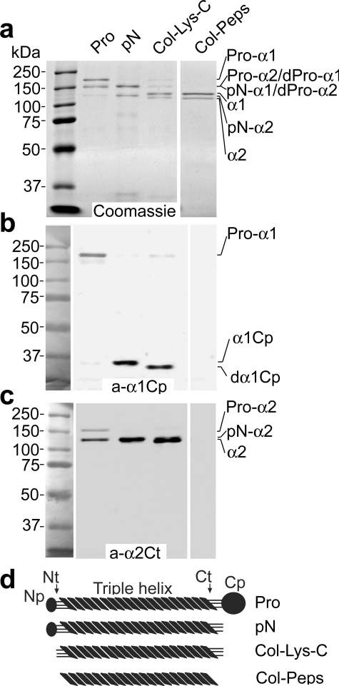
Analysis of specificity of epitope recognition by the anti-α1Cp
and anti-α2Ct antibodies. Panel a depicts intact
procollagen I and products of its digestion by BMP-1, Lys-C, or pepsin
separated in polyacrylamide gels and stained with Coomassie Blue. Specific
epitopes present in proteins represented in panel a were analyzed by
Western blot with the anti-α1Cp antibody (panel b) or with the
anti-α2Ct antibody (panel c). In panel d schematics of
domains in intact procollagen I or in products of its digestion by BMP-1,
Lys-C, or pepsin are presented. In addition, molecular mass markers are
presented. Pro, intact procollagen I; pN, procollagen
variant in which the C-terminal propeptide have been cleaved by BMP-1;
Col-Lys-C, collagen I variant, includes telopeptides, generated by
enzymatic cleavage of procollagen with Lys-C; Col-Peps, collagen I
variant, with telopeptides absent, generated by enzymatic cleavage of
procollagen I with pepsin; Pro-α1, pro-α1 chain
of procollagen I; Pro-α2, pro-α2 chain of
procollagen I; dPro-α1 and
dPro-α2, partially processed pro-α chains of
procollagen I; pN-α1 and
pN-α2, procollagen α chains in which the
C-terminal propeptides were cleaved by BMP-1; α1 and
α2, collagen I α chains; a-α1Cp
and a-α2Ct, antibodies against the C-terminal
propeptide of the α1 chain and the C-terminal telopeptide of the
α2 chain of procollagen I, respectively; α1Cp and
dα1Cp, C propeptide derived by cleavage of the
procollagen α1 chain with BMP-1 or Lys-C, respectively; Np, Nt,
Ct, and Cp, N-terminal propeptides, N-terminal telopeptides,
C-terminal telopeptides, and C-terminal propeptides of procollagen I,
respectively.
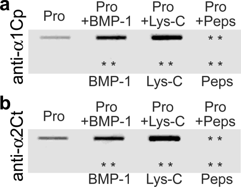
Slot blot analysis of the specificity of recognition of native epitopes
by the anti-α1Cp and anti-α2Ct antibodies. Panel a
shows results of assays of binding of the anti-α1Cp antibody to native
procollagen I and to the products of its cleavage with BMP-1, Lys-C, or
pepsin. Panel b depicts results of analyses of binding of the
anti-α2Ct antibody to the same proteins. In addition, both panels show
the lack of binding of analyzed antibodies to BMP-1, Lys-C, and pepsin.
Pro, procollagen I; + indicates samples digested with BMP-1, Lys-C,
or pepsin; asterisks indicate positions of proteins present on
nitrocellulose membranes but not detected by analyzed antibodies.
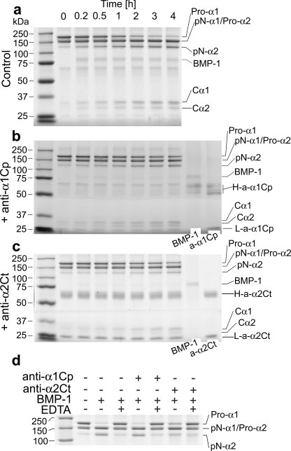
Kinetics of cleavage of procollagen I by BMP-1 in the presence of the
anti-α1Cp or the anti-α2Ct antibody. Panel a
illustrates kinetics of cleavage of procollagen I by BMP-1 in the absence of
antibodies, whereas panels b and c show kinetics of cleavage
of procollagen I in the presence of 1.2 μg/ml anti-α1Cp or the
anti-α2Ct antibody, respectively. Time points at which samples were
analyzed are indicated. In panels b and c, protein bands
corresponding to BMP-1 and to antibodies employed in this study are also
indicated. In addition, molecular mass markers are presented in the left
lanes of all panels. Panel d presents analysis of the
specificity of cleavage of procollagen I with BMP-1. Note that in the presence
of EDTA there was no cleavage of a procollagen substrate by BMP-1, thereby
indicating the absence of other active enzymes.
Pro-α1, pro-α1 chain of procollagen I;
Pro-α2, pro-α2 chain of procollagen I;
pN-α1 and pN-α2, procollagen
α chains in which the C-terminal propeptides were cleaved by BMP-1;
Cα1 and Cα2, C propeptides
cleaved off respective procollagen chains by BMP-1;
a-α1Cp and a-α2Ct, antibodies
against the C-terminal propeptide of the α1 chain and the C-terminal
telopeptide of the α2 chain of procollagen I, respectively;
H-a-α1Cp and L-a-α1Cp, heavy and light
chains of an anti-α1Cp immunoglobulin, respectively;
H-a-α2Ct and L-a-α2Ct, heavy
and light chains of an anti-α2Ct immunoglobulin, respectively; + and
– indicate the presence or the absence of selected compounds.
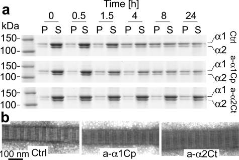
Kinetics of de novo formation of collagen fibrils.
Panel a represents analysis of collagen present in fibril and monomer
fractions at indicated time points. The upper row depicts control
samples, whereas the middle and lower rows show kinetics of
fibril formation in the presence of 180 μg/ml anti-α1Cp and
anti-α2Ct antibodies, respectively. In the left lane molecular
mass markers are indicated. Panel b demonstrates morphology of
individual fibrils formed in the presence or absence of tested antibodies.
P, pellet fractions that represents collagen fibrils; S,
supernatant fractions that represents collagen monomers; α1 and
α2, collagen I α chains; Ctrl, fibril formation
in the absence of antibodies; a-α1Cp and
a-α2Ct, fibril formation in the presence of antibodies
against the C-terminal propeptide of the α1 chain or the C-terminal
telopeptide of the α2 chain of procollagen I, respectively.
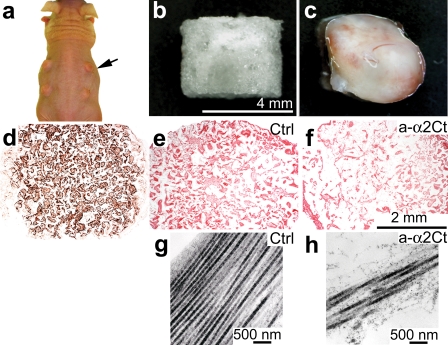
Analysis of morphology of keloid-like constructs formed subcutaneously
in nude mice. Panel a shows keloid-like constructs
(arrow) implanted under skin of a mouse. Panel b presents a
sponge-like scaffold that was employed to host human keloid-derived
fibroblasts. Panel c represents overall morphology of a keloid-like
construct harvested after 1 month of culture under skin of a nude mouse.
Panel d demonstrates immunostaining for human-specific vimentin to
confirm human origin of cells cultured in subcutaneous scaffolds, whereas
panels e and f present Sirius red staining of collagen
deposits. Panels g and h show electron microscopy images of
collagen fibrils present in sections of keloid-like constructs. Individual
panels show morphology of collagenous matrices formed in the absence
(e and g) or presence (f and h) of
inhibitory antibodies. Ctrl, keloid-like constructs cultured in the
absence of antibodies; a-α2Ct, keloid-like constructs
cultured in the presence of inhibitory antibodies against the C-terminal
telopeptide of the α2 chain of procollagen I. Scale bars are
included for specific sets of panels.
Similar articles
-
Asporin inhibits collagen matrix-mediated intercellular mechanocommunications between fibroblasts during keloid progression.FASEB J. 2021 Jul;35(7):e21705. doi: 10.1096/fj.202100111R. FASEB J. 2021. PMID: 34105826
-
Target-Specific Delivery of an Antibody That Blocks the Formation of Collagen Deposits in Skin and Lung.Monoclon Antib Immunodiagn Immunother. 2017 Oct;36(5):199-207. doi: 10.1089/mab.2017.0044. Epub 2017 Oct 3. Monoclon Antib Immunodiagn Immunother. 2017. PMID: 28972447 Free PMC article.
-
Testing the anti-fibrotic potential of the single-chain Fv antibody against the α2 C-terminal telopeptide of collagen I.Connect Tissue Res. 2014 Apr;55(2):115-22. doi: 10.3109/03008207.2013.862528. Epub 2014 Jan 10. Connect Tissue Res. 2014. PMID: 24195607 Free PMC article.
-
Meprin α and meprin β: Procollagen proteinases in health and disease.Matrix Biol. 2015 May-Jul;44-46:7-13. doi: 10.1016/j.matbio.2015.01.010. Epub 2015 Jan 21. Matrix Biol. 2015. PMID: 25617491 Review.
-
Developmental roles of the BMP1/TLD metalloproteinases.Birth Defects Res C Embryo Today. 2006 Mar;78(1):47-68. doi: 10.1002/bdrc.20060. Birth Defects Res C Embryo Today. 2006. PMID: 16622848 Review.
Cited by
-
Therapeutic Strategies to Overcome Fibrotic Barriers to Nanomedicine in the Pancreatic Tumor Microenvironment.Cancers (Basel). 2023 Jan 24;15(3):724. doi: 10.3390/cancers15030724. Cancers (Basel). 2023. PMID: 36765684 Free PMC article. Review.
-
Preparation and Characterization of Hydroxylated Recombinant Collagen by Incorporating Proline and Hydroxyproline in Proline-Deficient Escherichia coli.Bioengineering (Basel). 2024 Sep 27;11(10):975. doi: 10.3390/bioengineering11100975. Bioengineering (Basel). 2024. PMID: 39451351 Free PMC article.
-
Bone Mineralization in Electrospun-Based Bone Tissue Engineering.Polymers (Basel). 2022 May 23;14(10):2123. doi: 10.3390/polym14102123. Polymers (Basel). 2022. PMID: 35632005 Free PMC article. Review.
-
The impact of cholesterol deposits on the fibrillar architecture of the Achilles tendon in a rabbit model of hypercholesterolemia.J Orthop Surg Res. 2019 Jun 10;14(1):172. doi: 10.1186/s13018-019-1217-7. J Orthop Surg Res. 2019. PMID: 31182124 Free PMC article.
-
Mechanisms of reducing joint stiffness by blocking collagen fibrillogenesis in a rabbit model of posttraumatic arthrofibrosis.PLoS One. 2021 Sep 7;16(9):e0257147. doi: 10.1371/journal.pone.0257147. eCollection 2021. PLoS One. 2021. PMID: 34492074 Free PMC article.
References
-
- Prockop, D. J., and Kivirikko, K. I. (1995) Annu. Rev. Biochem. 64 403–434 - PubMed
-
- Colige, A., Vandenberghe, I., Thiry, M., Lambert, C. A., Van Beeumen, J., Li, S. W., Prockop, D. J., Lapiere, C. M., and Nusgens, B. V. (2002) J. Biol. Chem. 277 5756–5766 - PubMed
-
- Kessler, E., Takahara, K., Biniaminov, L., Brusel, M., and Greenspan, D. S. (1996) Science 271 360–362 - PubMed
-
- Addicks, E. M., Quigley, H. A., Green, W. R., and Robin, A. L. (1983) Arch. Ophthalmol. 101 795–798 - PubMed
Publication types
MeSH terms
Substances
Grants and funding
LinkOut - more resources
Full Text Sources
Other Literature Sources
Research Materials
Miscellaneous
