. 2008 Sep 19;283(38):25839-45.
doi: 10.1074/jbc.M802951200. Epub 2008 Jul 23.

Regulatory ATPase sites of cytoplasmic dynein affect processivity and force generation

Affiliations

Regulatory ATPase sites of cytoplasmic dynein affect processivity and force generation

Carol Cho et al. J Biol Chem. .

Abstract

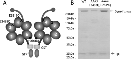
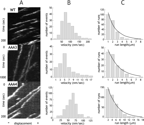
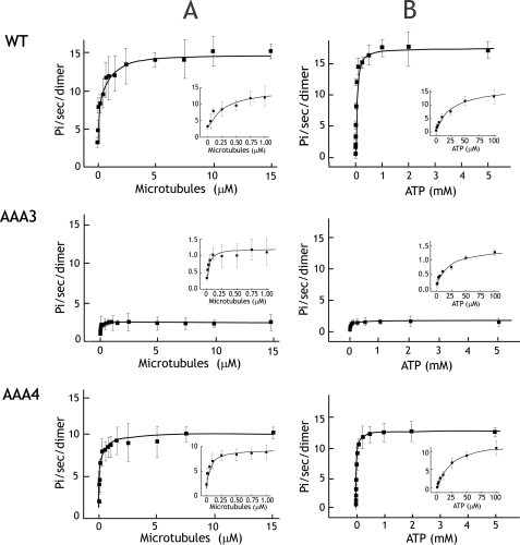
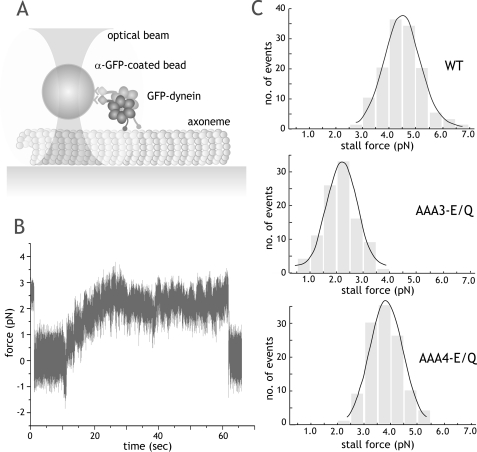
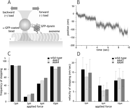
The heavy chain of cytoplasmic dynein contains four nucleotide-binding domains referred to as AAA1-AAA4, with the first domain (AAA1) being the main ATP hydrolytic site. Although previous studies have proposed regulatory roles for AAA3 and AAA4, the role of ATP hydrolysis at these sites remains elusive. Here, we have analyzed the single molecule motility properties of yeast cytoplasmic dynein mutants bearing mutations that prevent ATP hydrolysis at AAA3 or AAA4. Both mutants remain processive, but the AAA4 mutant exhibits a surprising increase in processivity due to its tighter affinity for microtubules. In addition to changes in motility characteristics, AAA3 and AAA4 mutants produce less maximal force than wild-type dynein. These results indicate that the nucleotide binding state at AAA3 and AAA4 can allosterically modulate microtubule binding affinity and affect dynein processivity and force production.


Figures

FIGURE 1.
FIGURE 1.
Construction and purification of dynein ATP hydrolysis mutants. A, a minimal S. cerevisiae cytoplasmic dynein that demonstrates processive motility was engineered as described previously (17). A glutathione S-transferase tag (GST) was incorporated at the NH2 terminus for the dimerization of the two heads of cytoplasmic dynein, whereas a HaloTag was fused to the COOH terminus for fluorescent labeling of the heads. In this paper, this construct is referred to as “wild-type dynein.” Highly conserved glutamate residues in the Walker B motif of domains AAA3 (E2488) and AAA4 (E2819) were mutated to glutamine to block ATP hydrolysis. B, Coomassie Blue-stained polyacrylamide gel of recombinant cytoplasmic dynein purified from S. cerevisiae by affinity purification. 330-kDa recombinant dynein is purified with minor amounts of 26-kDa IgG from the affinity matrix. WT, wild type.
FIGURE 2.
FIGURE 2.
Single molecule processivity of dynein ATP hydrolysis mutants. A, kymographs of single molecules of wild-type (WT) or ATP hydrolysis mutants. The x axis represents the length of an axoneme, and the y axis shows time. B, velocity histograms of wild-type and ATP hydrolysis mutants. The mean velocities ± S.D. are 73.9 ± 34.2 nm/s, 4.6 ± 3.7 nm/s, and 60.6 ± 18.9 nm/s (n = 221, 117, and 384) for wild type, AAA3-E/Q, and AAA4-E/Q, respectively. C, run length histograms of wild-type and ATP hydrolysis mutants are distributed in a single exponential decay. Run lengths were corrected for photobleaching and average axoneme length, and calculations for correct binning were performed as previously described (17). Run lengths (±S.E. as estimated by bootstrapping (17)) are 2.25 ± 0.14, 1.79 ± 0.18, and 4.38 ± 0.45 μm for wild type, AAA3-E/Q, and AAA4-E/Q, respectively.
FIGURE 3.
FIGURE 3.
Microtubule-stimulated ATPase activity of dynein ATP hydrolysis mutants. A, microtubule-stimulated ATPase activity of wild type and AAA mutants at 2 mm ATP. The insets show detailed views of microtubule-stimulated ATPase activity at low microtubule concentrations. Km,MT values for wild type (WT), AAA3-E/Q, and AAA4-E/Q are 0.31, 0.03, and 0.069 μm. respectively. B, the ATP dependence of microtubule-stimulated ATPase activity measured with 5 μm taxol-stabilized microtubules. Insets show detailed views of the curve at low ATP concentrations. Km,ATP values for wild type, AAA3-E/Q, and AAA4-E/Q are 25.2, 24.6, and 24.7 μm, respectively. Each dot represents the mean ± S.D. from three measurements of one preparation. Mean values from three preparations are presented in Table 1.
FIGURE 4.
FIGURE 4.
Stall force measurements of ATPase mutant dynein in the optical trap. A, schematic representation of the fixed optical trap setup used for stall force measurements in this paper. B, a representative trace of a single AAA4-E/Q dynein motor moving against force in a fixed optical trap at 1 mm ATP (trap stiffness (k) = 0.034 pN/nm). The trace shows a long stall event of ∼1 min, followed by release, which is typical of both wild-type and mutant dynein. C, stall force distributions of wild-type and ATPase mutant dyneins. Stall forces (mean ± S.D.) are 4.5 ± 1.3 pN (n = 132), 2.6 ± 1.2 pN (n = 100), and 3.7 ± 1.2 pN (n = 115) for wild type (WT), AAA3-E/Q, and AAA4-E/Q, respectively. Black lines, Gaussian fit of the data.
FIGURE 5.
FIGURE 5.
Nucleotide-independent movement of dynein induced by force. A, schematic of force-induced stepping experiments. Forward force is defined as the direction in which dynein normally moves (toward the microtubule minus-end). B, example trace of nucleotide-free, force-induced stepping for AAA4 mutants in a force feedback trap with 6 pN of backward load. k = 0.062 pN/nm. C, frequency of nucleotide-independent dynein movement after applying constant forward (–3 pN) or backward load (3, 6, and 10 pN). n > 25 molecules were tested at each force for each construct. Movement was scored within a 10-s time window of pulling on a dynein-coated bead attached to the microtubule. D, velocity of force-induced dynein movement with forward (–3 pN) or backward (6 and 10 pN) load (mean ± S.D.). Velocities at 3 pN backward load were not measured due to the small fraction of moving motors. GFP, green fluorescent protein.

References

    1. Vallee, R. B., Williams, J. C., Varma, D., and Barnhart, L. E. (2004) J. Neurobiol. 58 189–200 - PubMed
    1. Vale, R. D. (2000) J. Cell Biol. 150 F13–F19 - PMC - PubMed
    1. Ogura, T., and Wilkinson, A. J. (2001) Genes Cells 6 575–597 - PubMed
    1. Neuwald, A. F., Aravind, L., Spouge, J. L., and Koonin, E. V. (1999) Genome Res. 9 27–43 - PubMed
    1. Erzberger, J. P., and Berger, J. M. (2006) Annu. Rev. Biophys. Biomol. Struct. 35 93–114 - PubMed

Publication types

LinkOut - more resources