

Review
doi: 10.1182/blood-2007-09-077438.
Human natural killer cells
Affiliations
- PMID: 18650461
- PMCID: PMC2481557
- DOI: 10.1182/blood-2007-09-077438
Item in Clipboard
Review
Human natural killer cells
Blood.
.
Abstract
Natural killer (NK) cells were discovered more than 30 years ago. NK cells are large granular lymphocytes that belong to the innate immune system because unlike T or B lymphocytes of the adaptive or antigen-specific immune system, NK cells do not rearrange T-cell receptor or immunoglobulin genes from their germline configuration. During the past 2 decades there has been a substantial gain in our understanding of what and how NK-cells "see," lending important insights into their functions and purpose in normal immune surveillance. The most recent discoveries in NK-cell receptor biology have fueled translational research that has led to remarkable results in treating human malignancy.
Figures

Michael A. Caligiuri
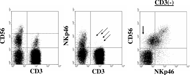
Human NK cells are CD3−CD56+NKp46+. Shown is a representative example of immunophenotypic analysis from a healthy donor when gating on peripheral blood lymphocytes. NK cells lack expression of CD3 and coexpress CD56 and NKp46. The left and middle dot plots were gated on total lymphocytes using forward scatter versus side scatter parameters. The dashed line in the left dot plot separates CD56dim from CD56bright NK cells. The arrows in the middle plot indicate a few CD3+ cells that coexpress NKp46. The right dot plot is gated on CD3− lymphocytes and demonstrates the 2 major NK-cell populations in the blood: CD56brightNKp46bright and CD56dimNKp46dim. The arrow in the first quadrant of the right dot plot highlights a few CD56dim NK cells that likely lack expression of NKp46.
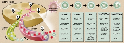
Model of human NK-cell development. (1) Bone marrow–derived CD34+CD45RA+ HPCs circulate in the blood and (2) extravasate across lymph node high endothelial venules to enter the parafollicular space. There, (3) pro-NK cells are activated to progress through distinct stages of maturation (far right) to create both CD56bright and CD56dim NK cells. Maturing CD56dim NK cells return to the circulation via the efferent lymph (4), whereas some CD56bright NK cells remain within the secondary lymphoid tissue to interact with DCs (5).,,, Illustration by Debra T. Dartez.
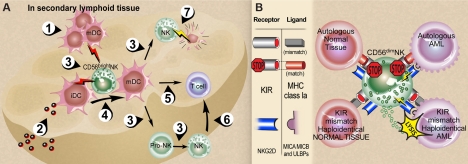
CD56bright and CD56dim NK-cell interactions. (A) NK-DC interactions in secondary lymphoid tissue (SLT). (1) Activated mature DCs (mDCs) enter SLT from periphery or (2) immature DCs (iDCs) receive pathogens within SLT. Each express and/or secrete a variety of cytokines (3) that are required for NK-cell maturation and survival (eg, DC IL-15) and NK cell proinflammatory cytokine production (eg, DC IL-12 in combination with DC IL-1, IL-15, IL-18). Activated CD56bright NK cells in turn secrete TNF-α and GM-CSF that contribute to DC maturation, (4) and IFN-γ that contributes to DC activation and thus indirectly to antigen-specific T-cell priming (5). NK-cell IFN-γ also contributes directly to T-cell priming (6). NK cells can kill immature autologous DCs (7) via NKp30, which may assist in editing out hyporesponsive DCs or by limiting T-cell priming., (B) Summary of NK-cell recognition. The functional consequences of NK-cell receptor recognition depend on the integration of both inhibition and activation signals received in response to engagement of target cell ligands., Upper left: normal autologous tissues are not attacked because the predominant signal is recognition of self-MHC class Ia ligands by inhibitory KIRs (and other inhibitory receptors such as NKG2A/CD94 recognizing their ligands, not shown) in the absence of ligands for activating NK receptors. Upper right: Malignant autologous tumors such as acute myeloid leukemia (AML) have high-density surface expression of classical MHC class Ia and nonclassical MHC class I that that bind to KIR and NKG2A/CD94, respectively, and dominate over engagement of NK-cell activation receptors with their cognate ligands. Lower left: Normal allogeneic host tissue presumably lacks ligands that engage dominant activating NK receptors such as NKG2D and NCR, despite a mismatch of donor NK KIR with host MHC class Ia as well as donor NKG2A/CD94 and host HLA-E (not shown). Lower right: A mismatch of donor NK KIR and host MHC class Ia in the presence of ligand-engaged NKG2D, NCR, and other NK activation receptors likely contributes the dominant NK response of target cell lysis.,, Illustration by Debra T. Dartez.
References
-
- Cooper MD, Alder MN. The evolution of adaptive immune systems. Cell. 2006;124:815–822. - PubMed
-
- Orange JS. Human natural killer cell deficiencies. Curr Opin Allergy Clin Immunol. 2006;6:399–409. - PubMed
-
- Herberman RB, Nunn ME, Holden HT, Lavrin DH. Natural cytotoxic reactivity of mouse lymphoid cells against syngeneic and allogeneic tumors, II: characterization of effector cells. Int J Cancer. 1975;16:230–239. - PubMed
-
- Kiessling R, Klein E, Pross H, Wigzell H. “Natural” killer cells in the mouse, II: cytotoxic cells with specificity for mouse Moloney leukemia cells: characteristics of the killer cell. Eur J Immunol. 1975;5:117–121. - PubMed
-
- Lanier LL, Phillips JH, Hackett J, Jr, Tutt M, Kumar V. Natural killer cells: definition of a cell type rather than a function. J Immunol. 1986;137:2735–2739. - PubMed
Publication types
MeSH terms
Substances
Grants and funding
LinkOut - more resources
Full Text Sources
Other Literature Sources
Miscellaneous
