

Bioengineered dental tissues grown in the rat jaw
- PMID: 18650546
- PMCID: PMC3024580
- DOI: 10.1177/154405910808700811
Bioengineered dental tissues grown in the rat jaw
Abstract
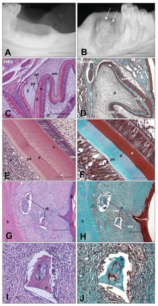
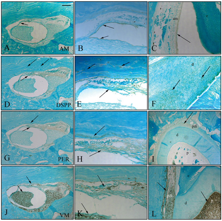
Our long-term objective is to develop methods to form, in the jaw, bioengineered replacement teeth that exhibit physical properties and functions similar to those of natural teeth. Our results show that cultured rat tooth bud cells, seeded onto biodegradable scaffolds, implanted into the jaws of adult rat hosts and grown for 12 weeks, formed small, organized, bioengineered tooth crowns, containing dentin, enamel, pulp, and periodontal ligament tissues, similar to identical cell-seeded scaffolds implanted and grown in the omentum. Radiographic, histological, and immunohistochemical analyses showed that bioengineered teeth consisted of organized dentin, enamel, and pulp tissues. This study advances practical applications for dental tissue engineering by demonstrating that bioengineered tooth tissues can be regenerated at the site of previously lost teeth, and supports the use of tissue engineering strategies in humans, to regenerate previously lost and/or missing teeth. The results presented in this report support the feasibility of bioengineered replacement tooth formation in the jaw.
Figures
Similar articles
-
Bioengineered teeth from cultured rat tooth bud cells.J Dent Res. 2004 Jul;83(7):523-8. doi: 10.1177/154405910408300703. J Dent Res. 2004. PMID: 15218040
-
Tissue-engineered hybrid tooth and bone.Tissue Eng. 2005 Sep-Oct;11(9-10):1599-610. doi: 10.1089/ten.2005.11.1599. Tissue Eng. 2005. PMID: 16259613
-
Reconstructing mandibular defects using autologous tissue-engineered tooth and bone constructs.J Oral Maxillofac Surg. 2009 Feb;67(2):335-47. doi: 10.1016/j.joms.2008.09.002. J Oral Maxillofac Surg. 2009. PMID: 19138608
-
Bioengineered teeth from tooth bud cells.Dent Clin North Am. 2006 Apr;50(2):191-203, viii. doi: 10.1016/j.cden.2005.11.005. Dent Clin North Am. 2006. PMID: 16530057 Review.
-
Tooth Repair and Regeneration: Potential of Dental Stem Cells.Trends Mol Med. 2021 May;27(5):501-511. doi: 10.1016/j.molmed.2021.02.005. Epub 2021 Mar 26. Trends Mol Med. 2021. PMID: 33781688 Free PMC article. Review.
Cited by
-
Tooth tissue engineering: the influence of hydrophilic surface on nanocrystalline diamond films for human dental stem cells.Tissue Eng Part A. 2013 Dec;19(23-24):2537-43. doi: 10.1089/ten.TEA.2012.0628. Epub 2013 Oct 12. Tissue Eng Part A. 2013. PMID: 23863195 Free PMC article.
-
Craniofacial Tissue Engineering.Cold Spring Harb Perspect Med. 2018 Jan 2;8(1):a025775. doi: 10.1101/cshperspect.a025775. Cold Spring Harb Perspect Med. 2018. PMID: 28348178 Free PMC article. Review.
-
Tooth Tissue Engineering: The Importance of Blood Products as a Supplement in Tissue Culture Medium for Human Pulp Dental Stem Cells.Tissue Eng Part A. 2015 Nov;21(21-22):2639-48. doi: 10.1089/ten.TEA.2014.0617. Tissue Eng Part A. 2015. PMID: 26414682 Free PMC article.
-
"Opening" the mesenchymal stem cell tool box.Eur J Dent. 2009 Jul;3(3):240-9. Eur J Dent. 2009. PMID: 19756201 Free PMC article.
-
Calcium orthophosphates in dentistry.J Mater Sci Mater Med. 2013 Jun;24(6):1335-63. doi: 10.1007/s10856-013-4898-1. Epub 2013 Mar 7. J Mater Sci Mater Med. 2013. PMID: 23468163 Review.
References
-
- Braut A, Kollar EJ, Mina M. Analysis of the odontogenic and osteogenic potentials of dental pulp in vivo using a col1a1-2.3-GFP transgene. Int J Dev Biol. 2003;47:281–292. - PubMed
-
- Cai J, Cho SW, Kim JY, Lee MJ, Cha YG, Jung HS. Patterning the size and number of tooth and its cusps. Dev Biol. 2007;304:499–507. - PubMed
-
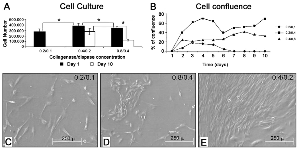
- Cortes O, Garcia C, Perez L, Boj J, Alcaina A. Pulp cell cultures obtained with two different methods for in vitro cytotoxicity tests. Eur Arch Paediatr Dent. 2006;7:96–99. - PubMed
-
- Duailibi MT, Duailibi SE, Young CS, Vacanti JP, Bartlett JD, Yelick PC. Bioengineered teeth from cultured rat tooth bud cells. J Dent Res. 2004;83:523–528. - PubMed
-
- Duailibi SE, Duailibi MT, Vacanti JP, Yelick PC. Prospects for tooth regeneration. Periodontol 2000. 2006;41:177–187. - PubMed
Publication types
MeSH terms
Substances
Grants and funding
LinkOut - more resources
Full Text Sources
Other Literature Sources
