. 2008 Aug 28;454(7208):1142-6.
doi: 10.1038/nature07163. Epub 2008 Jul 23.

Cell-specific ATP7A transport sustains copper-dependent tyrosinase activity in melanosomes

Affiliations

Cell-specific ATP7A transport sustains copper-dependent tyrosinase activity in melanosomes

Subba Rao Gangi Setty et al. Nature. .

Abstract

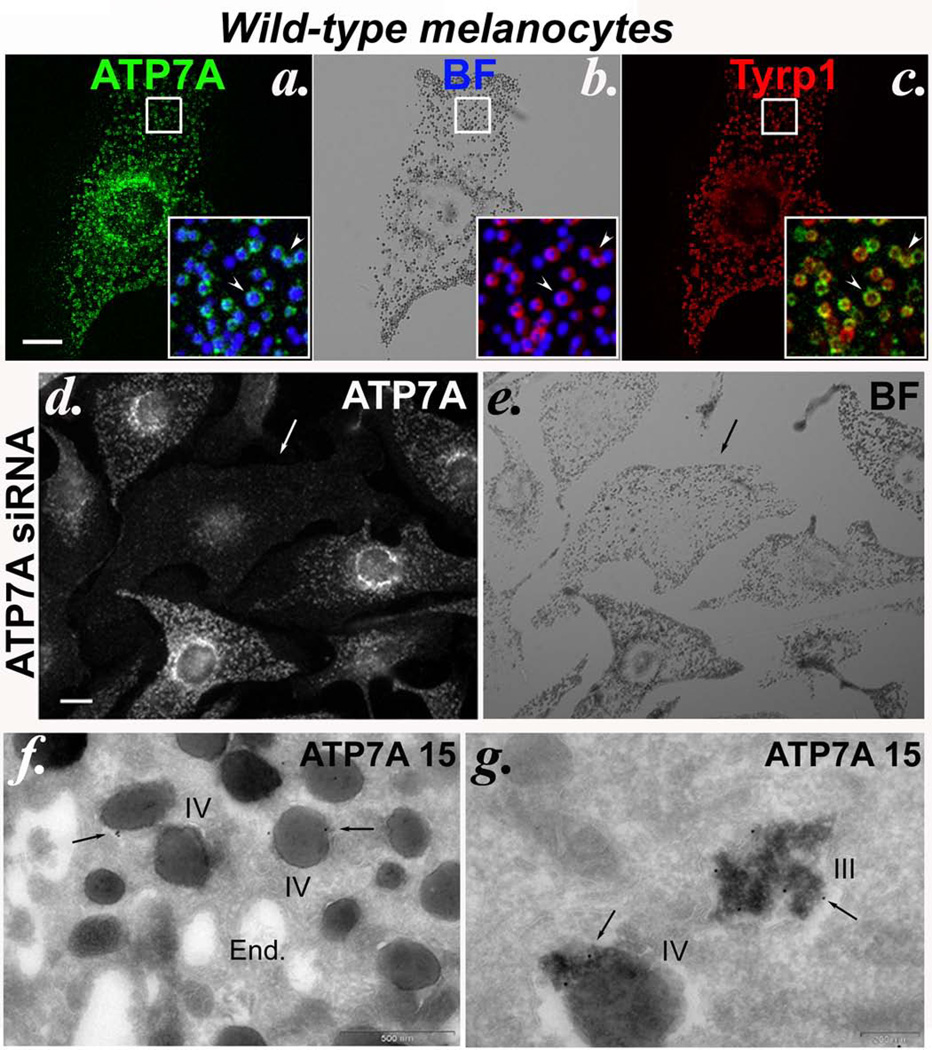
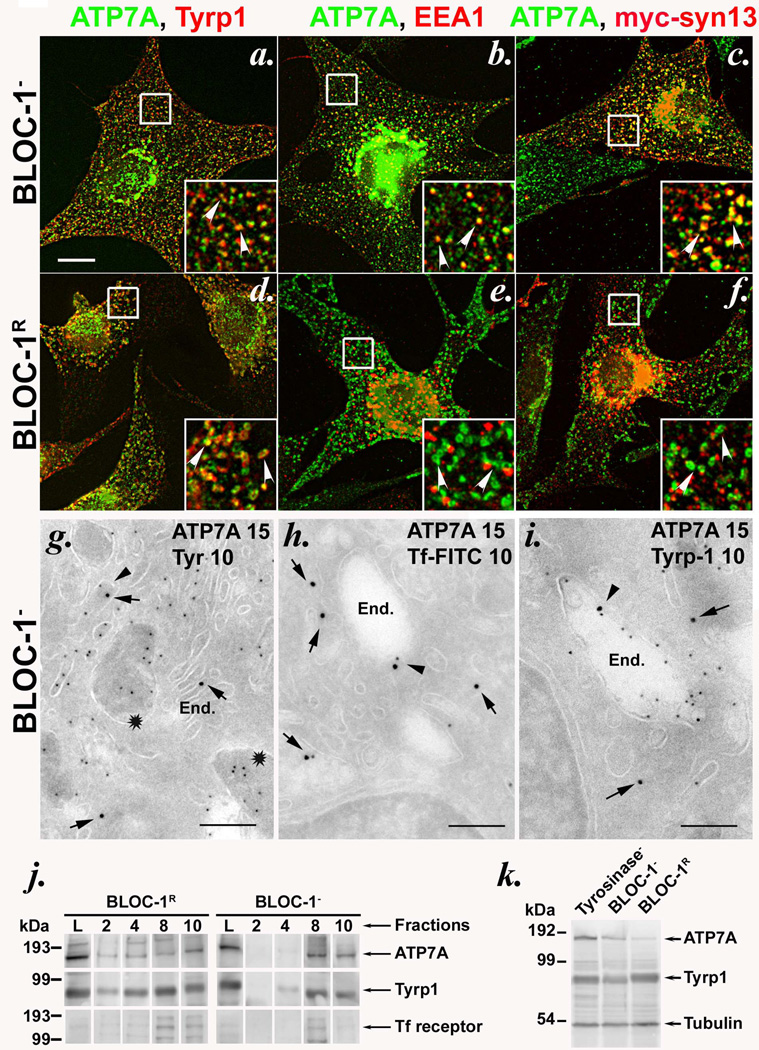
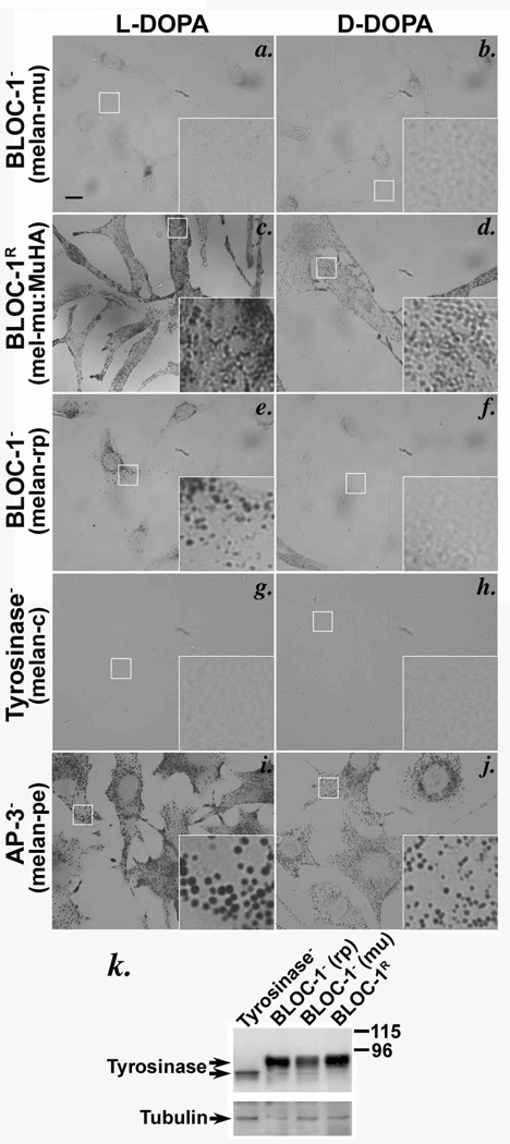
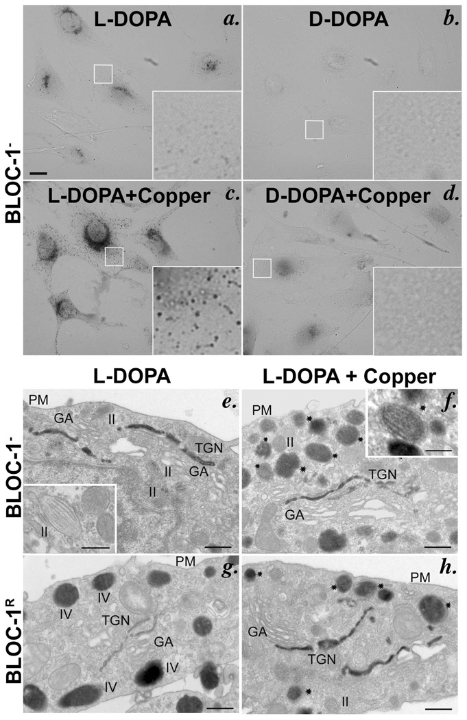
Copper is a cofactor for many cellular enzymes and transporters. It can be loaded onto secreted and endomembrane cuproproteins by translocation from the cytosol into membrane-bound organelles by ATP7A or ATP7B transporters, the genes for which are mutated in the copper imbalance syndromes Menkes disease and Wilson disease, respectively. Endomembrane cuproproteins are thought to incorporate copper stably on transit through the trans-Golgi network, in which ATP7A accumulates by dynamic cycling through early endocytic compartments. Here we show that the pigment-cell-specific cuproenzyme tyrosinase acquires copper only transiently and inefficiently within the trans-Golgi network of mouse melanocytes. To catalyse melanin synthesis, tyrosinase is subsequently reloaded with copper within specialized organelles called melanosomes. Copper is supplied to melanosomes by ATP7A, a cohort of which localizes to melanosomes in a biogenesis of lysosome-related organelles complex-1 (BLOC-1)-dependent manner. These results indicate that cell-type-specific localization of a metal transporter is required to sustain metallation of an endomembrane cuproenzyme, providing a mechanism for exquisite spatial control of metalloenzyme activity. Moreover, because BLOC-1 subunits are mutated in subtypes of the genetic disease Hermansky-Pudlak syndrome, these results also show that defects in copper transporter localization contribute to hypopigmentation, and hence perhaps other systemic defects, in Hermansky-Pudlak syndrome.


Figures

Figure 1
Figure 1. Copper transporter ATP7A localizes to melanosomes in wild-type melanocytes
(a–c) Fixed melan-a (wild-type) cells were analyzed by IFM with antibodies to ATP7A and Tyrp1. The corresponding bright field (BF) image shows pigmented melanosomes. Insets, 3X-magified overlays of the boxed regions comparing ATP7A to melanosomes (pseudocolored blue from an inverted BF image, left), melanosomes to Tyrp1 (middle) and ATP7A to Tyrp1 (right). Arrowheads, colocalized ATP7A, melanosomes and Tyrp1. Bar, 10 µm. (d, e) Wild-type melan-Ink4a melanocytes were transduced with siRNA to ATP7A (ATP7A-3 siRNA) and analyzed 48 h later by IFM for ATP7A and BF microscopy. Arrow, a cell in which labeling for ATP7A on pigment granules is lost. Bar, 10 µm. (f, g) Ultrathin cryosections of wild-type melan-a cells were immunogold labeled with anti-ATP7A antibody and analyzed by electron microscopy. Arrows indicate labeling of stage III and IV melanosomes (III and IV), predominantly on the internal membranes within them. See also Supplementary Figure S4. End., endosomes. Bars, 500 nm (f) and 200 nm (g).
Figure 2
Figure 2. ATP7A is mislocalized to early endosomes in BLOC-1-deficient melanocytes
(a–f) IFM analysis of BLOC-1 (melan-mu, a–c) and BLOC-1R (melan-mu: MuHA, d–f) melanocytes labeled with antibodies to ATP7A and either Tyrp1 (left), EEA1 (middle) or transiently expressed myc epitope-tagged Syntaxin 13 (myc-syn13, right). Insets, boxed regions magnified 3X. Arrowheads, ATP7A colocalized with the indicated marker. Bar, 10 µm. Note that Tyrp1 labels melanosomes in BLOC-1R cells but early endosomes in BLOC-1 cells; EEA1 and syn13 label early endosomes in both cell types. (g–i) Ultrathin cryosections of BLOC-1 melan-mu cells were immunogold labeled with antibodies to ATP7A (15 nm gold) and tyrosinase (g), internalized Tf (h) or Tyrp1 (i)(10 nm gold) and analyzed by electron microscopy. Arrows indicate labeling of ATP7A and arrowheads indicate ATP7A with Tf or Tyrp1 on endosomes. Stars in (g) indicate striated melanosomes labeled for tyrosinase but not ATP7A. End., endosomes. Bars, 500 nm. See also Supplementary Figure S4. (j) Subcellular fractionation of BLOC-1 (melan-mu) and BLOC-1R (melan-mu:MuHA) cells on sucrose step gradients. Eluted fractions (2, 4, 8 and 10), collected from bottom to top, and lysates (L, input loading control) were probed by immunoblot with antibodies to ATP7A, Tyrp1 and Tf receptor (to label early endosomes). Left, molecular weight markers (in kDa). Arrows, relevant bands. Note the bands for ATP7A and Tyrp1, but not Tf receptor (middle band), in pigment-containing fractions 2 and 4 from melan-mu:MuHA but not melan-mu cells. (k) Whole cell lysates from tyrosinase-deficient (Tyrosinase-, melan-c), BLOC-1 (melan-mu) and BLOC-1R (melan-mu:MuHA) were fractionated by SDS-PAGE and immunoblotted with antibodies to the ATP7A, Tyrp1 and tubulin. Left, molecular weight markers (in kDa). Arrows, relevant bands.
Figure 3
Figure 3. Tyrosinase is present but inactive in BLOC-1-deficient melanocytes
(a–j) Bright field microscopy analysis of BLOC-1 (melan-mu, a, b; melan-rp, e, f), BLOC-1R (melan-mu:MuHA, c, d), AP-3-deficient (melan-pe, i, j) and tyrosinase mutant (melan-c, g, h) mouse melanocytes treated with L- or D-DOPA for 3 h. Tyrosinase activity is indicated by melanin deposition in the presence of L-DOPA but not D-DOPA. Insets, boxed regions magnified 5X. Bars, 10 µm. Note melanin deposits were not observed in melan-mu (a) or control melan-c cells (g), and modest deposits were observed in melan-rp cells in which BLOC-1 is partially assembled (e). (k) Whole cell lysates of mouse melanocytes were fractionated by SDS-PAGE and immunoblotted with anti-tyrosinase antibody. Blots were reprobed with anti-tubulin antibody as a loading control. Right, molecular weight markers (kDa). Arrows, relevant bands. Note that tyrosinase protein levels are only mildly reduced in BLOC-1 melan-mu cells relative to BLOC-1R melan-mu: MuHA cells.
Figure 4
Figure 4. Copper restores in vitro tyrosinase activity in melanosomes of BLOC-1-deficient melanocytes
DOPA cytochemistry of BLOC-1 melan-mu (a–f) or BLOC-1R melan-mu:MuHA (g, h) cells in the absence or presence (+ Copper) of 20 µM copper sulfate in the reaction buffer. (a–d) Bright field microscopy analysis of cells treated for 4 h as indicated. Insets, boxed regions magnified 10X. Bars, 10 µm. Note the increased melanin deposits in BLOC-1 cells in the presence (c) compared to the absence (a) of copper. (e–h) EM analysis of thin sections of cells treated for 2 h. Note melanin deposits in the trans-most Golgi cisternae, but not in striated melanosomes, of BLOC-1 melan-mu cells in the absence of excess copper (e; inset), and the additional deposition of melanin in striated melanosomes in the presence of copper (f, stars; inset). GA, Golgi apparatus; TGN, trans-Golgi network; PM, Plasma membrane; II, III, IV, melanosome stages II and IV. Bars, 500 nm and inset, 200 nm.

References

    1. Thiele DJ. Integrating trace element metabolism from the cell to the whole organism. J. Nutr. 2003;133:1579S–1580S. - PubMed
    1. Mercer JF. The molecular basis of copper-transport diseases. Trends Mol. Med. 2001;7:64–69. - PubMed
    1. Yamaguchi Y, Heiny ME, Suzuki M, et al. Biochemical characterization and intracellular localization of the Menkes disease protein. Proc. Natl. Acad. Sci. U.S.A. 1996;93:14030–14035. - PMC - PubMed
    1. Harris ED. Cellular copper transport and metabolism. Annu. Rev. Nutr. 2000;20:291–310. - PubMed
    1. Lutsenko S, Barnes NL, Bartee MY, et al. Function and regulation of human copper-transporting ATPases. Physiol. Rev. 2007;87:1011–1046. - PubMed

Publication types

MeSH terms