Review
. 2008 Jul 24;454(7203):455-62.
doi: 10.1038/nature07203.

From endoplasmic-reticulum stress to the inflammatory response

Affiliations
Review

From endoplasmic-reticulum stress to the inflammatory response

Kezhong Zhang et al. Nature. .

Abstract

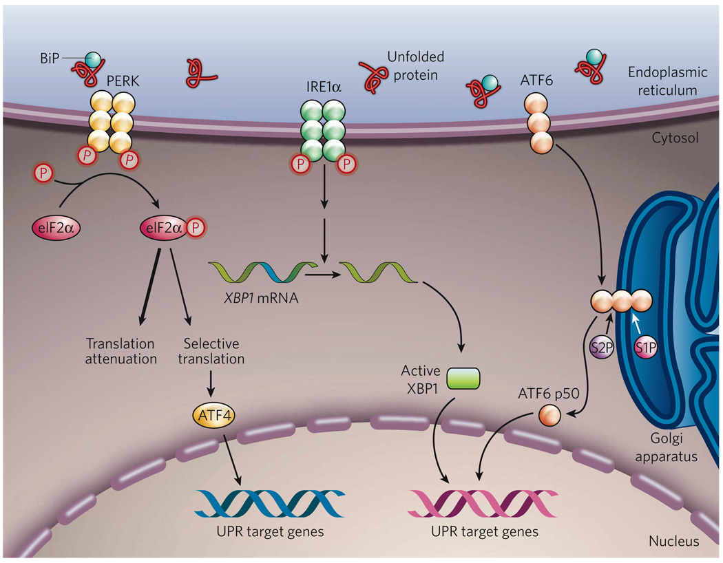
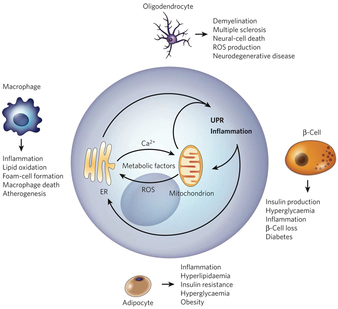
The endoplasmic reticulum is responsible for much of a cell's protein synthesis and folding, but it also has an important role in sensing cellular stress. Recently, it has been shown that the endoplasmic reticulum mediates a specific set of intracellular signalling pathways in response to the accumulation of unfolded or misfolded proteins, and these pathways are collectively known as the unfolded-protein response. New observations suggest that the unfolded-protein response can initiate inflammation, and the coupling of these responses in specialized cells and tissues is now thought to be fundamental in the pathogenesis of inflammatory diseases. The knowledge gained from this emerging field will aid in the development of therapies for modulating cellular stress and inflammation.


Figures

Figure 1
Figure 1. The mammalian UPR pathways
In non-stressed cells (not shown), the ER chaperone BiP binds to the luminal domains of the ER-stress sensors IRE1α, PERK and ATF6, maintaining these proteins in an inactive state. During ER stress (shown), BiP preferentially binds to unfolded or misfolded proteins, thus driving the equilibrium of BiP binding away from IRE1α, PERK and ATF6. These three proteins are the initiators of the three main signalling cascades of the UPR. The release of BiP results in the activation of PERK, through PERK homodimerization and trans-autophosphorylation. Activated PERK then phosphorylates the translation-initiation factor eIF2α, reducing the overall frequency of messenger RNA translation initiation. However, selected mRNAs, such as ATF4 mRNA, are preferentially translated in the presence of phosphorylated eIF2α. ATF4 activates the transcription of UPR target genes encoding factors involved in amino-acid biosynthesis, the antioxidative-stress response and apoptosis. The release of BiP also allows IRE1α to dimerize, activating its protein-kinase activity (through autophosphorylation) and its endoribonuclease activity. IRE1α then removes a 26-base intron from XBP1 mRNA. The spliced XBP1 mRNA encodes a potent transcription factor that translocates to the nucleus, activating the expression of UPR target genes. The release of BiP from ATF6 allows ATF6 to translocate to the Golgi apparatus, where it is cleaved by the proteases S1P and S2P, yielding an active cytosolic ATF6 fragment (ATF6 p50). This fragment migrates to the nucleus, activating the transcription of UPR target genes. S1P, site-1 protease; S2P, site-2 protease; XBP1, X-box-binding protein 1.
Figure 2
Figure 2. Oxidative protein folding
The formation of disulphide bonds in proteins in the ER is driven by the enzymes PDI and ERO1. ERO1 operates in association with the flavin FAD, which is synthesized in the cytosol but can readily enter the ER lumen. PDI accepts electrons (e) from protein-folding substrates, thereby oxidizing the thiol (SH) groups in the protein’s cysteine residues and resulting in the formation of disulphide bonds. ERO1 uses an FAD-dependent reaction to transfer electrons from PDI to molecular oxygen (O2), resulting in the production of ROS in the form of hydrogen peroxide (H2O2). Reduced glutathione (GSH) can assist in disulphide-bond reduction, which occurs when there is a overload of proteins to fold or an accumulation of misfolded proteins, and results in the production of oxidized glutathione (GSSG). In addition, reduced PDI can mediate a reduction of mispaired thiol groups in oxidized protein-folding substrates, functioning as an isomerase. Because the activity of ERO1 is modulated by the amount of FAD in the ER, disulphide-bond formation is linked to the nutritional and/or metabolic status of the cell.
Figure 3
Figure 3. Proposed models for UPR-mediated JNK and NF-κB activation
In response to ER stress, PERK mediates a general repression of mRNA translation by phosphorylating eIF2α. Because IκB has a shorter half-life than NF-κB, PERK-mediated translational attenuation shifts the ratio of IκB to NF-κB, thereby freeing NF-κB to translocate to the nucleus. In addition, in response to ER stress, the cytoplasmic domain of phosphorylated IRE1α can recruit tumour-necrosis factor-α (TNF-α)-receptor-associated factor 2 (TRAF2). The IRE1α–TRAF2 complex interacts with JNK and/or IκB kinase (IKK), activating these protein kinases. Activated JNK phosphorylates the transcription factor activator protein 1 (AP1). Activated IKK phosphorylates IκB, initiating the degradation of IκB and thereby leading to NF-κB activation. Activated NF-κB and AP1 then migrate to the nucleus, where they induce the transcription of genes involved in the inflammatory response.
Figure 4
Figure 4. The ER-stress-induced acute-phase response
When inflammatory cytokines, such as TNF-α, IL-1β and IL-6, are present in the extracellular environment, the gene encoding CREBH is transcribed (not shown). CREBH, similar to ATF6, is a bZIP-containing transcription factor that is localized to the ER membrane. CREBH, however, is mainly expressed by hepatocytes, whereas ATF6 is expressed by all cell types. In conditions of ER stress, such as those caused by inflammatory cytokines or the bacterial component lipopolysaccharide (LPS), CREBH translocates to the Golgi apparatus, where it is cleaved by the proteases S1P and S2P, releasing a cytosolic fragment. ER stress also activates the UPR sensor ATF6 by regulated intramembrane proteolysis. Activated CREBH and ATF6 can then form homodimers or heterodimers and migrate to the nucleus, where they activate the transcription of the genes encoding serum amyloid P component and C-reactive protein, which mediate the acute-phase response.
Figure 5
Figure 5. The role of calcium and ROS in the UPR and inflammation
Protein folding is an oxidative process that generates ROS. ROS can target chaperones (not shown) and ER-based calcium (Ca2+) channels, leading to the release of calcium from the ER into the cytosol and ER-stress signalling. Calcium released from the ER is concentrated in the inner matrix of the mitochondria, where it disrupts the electron-transport chain, thereby leading to the production of more ROS. These mitochondrially produced ROS can further exacerbate calcium release from the ER, resulting in the accumulation of ROS to a toxic level. Furthermore, perturbation of ER calcium homeostasis can disrupt the protein-folding process in the ER, which, in turn, causes ER stress, induces the UPR and generates more ROS.
Figure 6
Figure 6. The ‘ER-stress–inflammation’ loop in specialized cells
In specialized cells that secrete large amounts of protein — such as macrophages, adipocytes, β-cells and oligodendrocytes — the UPR and inflammatory-response signalling can be triggered by a chronic excess of extracellular and/or intracellular metabolic factors, such as lipids, glucose, cytokines, hormones, non-esterified fatty acids and neurotransmitters. More specifically, such metabolic factors stimulate protein synthesis, calcium signalling and ROS production by targeting the mitochondria and the ER in these cells (Fig. 5). The increased protein-folding demand and the signalling involving calcium and ROS induce the UPR and inflammatory-response signalling, leading to the transcription of genes whose products mount a broader inflammatory response. An excess of metabolic factors can further boost the UPR and inflammation, contributing to impaired lipid and glucose metabolism, insulin resistance and apoptosis. This forward ER-stress– inflammation loop could also further promote inflammatory stress signalling and contribute to the metabolic deterioration that is associated with atherosclerosis, obesity, type 2 diabetes and neurodegenerative diseases, depending on the cell type involved.

References

    1. Charo IF, Ransohoff RM. The many roles of chemokines and chemokine receptors in inflammation. N. Engl. J. Med. 2006;354:610–621. - PubMed
    1. Hotamisligil GS. Inflammation and metabolic disorders. Nature. 2006;444:860–867. - PubMed
    1. Hansson GK, Libby P. The immune response in atherosclerosis: a double-edged sword. Nature Rev. Immunol. 2006;6:508–519. - PubMed
    1. Kaufman RJ. Stress signaling from the lumen of the endoplasmic reticulum: coordination of gene transcriptional and translational controls. Genes Dev. 1999;13:1211–1233. - PubMed
    1. Ron D, Walter P. Signal integration in the endoplasmic reticulum unfolded protein response. Nature Rev. Mol. Cell Biol. 2007;8:519–529. - PubMed

Publication types

Substances