. 2008 Aug 6;27(15):2031-42.
doi: 10.1038/emboj.2008.139. Epub 2008 Jul 24.

Reconstituted membrane fusion requires regulatory lipids, SNAREs and synergistic SNARE chaperones

Affiliations

Reconstituted membrane fusion requires regulatory lipids, SNAREs and synergistic SNARE chaperones

Joji Mima et al. EMBO J. .

Abstract

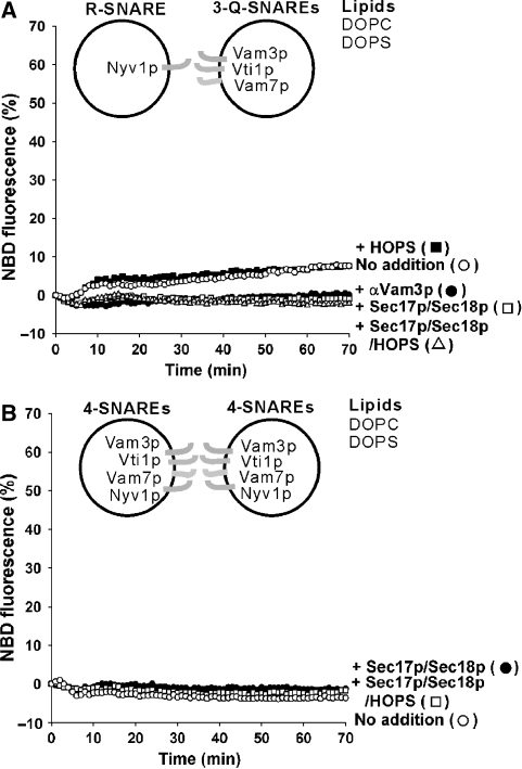
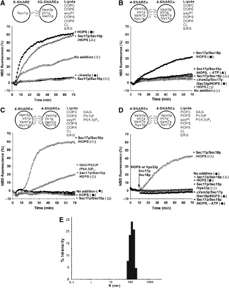
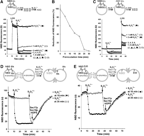
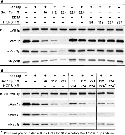
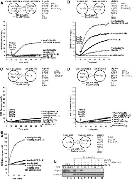
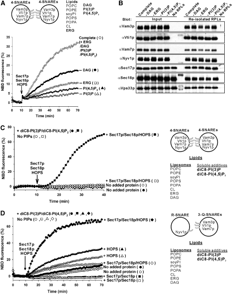
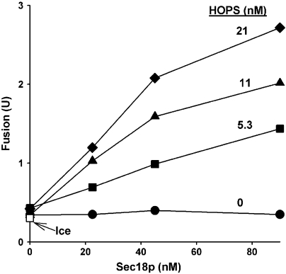
The homotypic fusion of yeast vacuoles, each with 3Q- and 1R-SNARE, requires SNARE chaperones (Sec17p/Sec18p and HOPS) and regulatory lipids (sterol, diacylglycerol and phosphoinositides). Pairs of liposomes of phosphatidylcholine/phosphatidylserine, bearing three vacuolar Q-SNAREs on one and the R-SNARE on the other, undergo slow lipid mixing, but this is unaffected by HOPS and inhibited by Sec17p/Sec18p. To study these essential fusion components, we reconstituted proteoliposomes of a more physiological composition, bearing vacuolar lipids and all four vacuolar SNAREs. Their fusion requires Sec17p/Sec18p and HOPS, and each regulatory lipid is important for rapid fusion. Although SNAREs can cause both fusion and lysis, fusion of these proteoliposomes with Sec17p/Sec18p and HOPS is not accompanied by lysis. Sec17p/Sec18p, which disassemble SNARE complexes, and HOPS, which promotes and proofreads SNARE assembly, act synergistically to form fusion-competent SNARE complexes, and this synergy requires phosphoinositides. This is the first chemically defined model of the physiological interactions of these conserved fusion catalysts.


Figures

Figure 1
Figure 1
SNARE-mediated lipid mixing of PC/PS liposomes. (A) Lipid mixing between PC/PS liposomes with vacuolar 3Q-SNAREs (270–560 nM) or R-SNARE (94 nM). (B) Lipid mixing between PC/PS liposomes bearing all the four SNAREs (480–700 nM). Sec17p (400 nM), Sec18p (400 nM) and HOPS (10 nM) were added where indicated. All data were from one experiment and are representative of five independent experiments.
Figure 2
Figure 2
Lipid mixing between SNARE proteoliposomes with vacuolar lipids, regulatory lipids, Sec17p/Sec18p and HOPS. (A) Effect of Sec17p (400 nM), Sec18p (400 nM) and HOPS (10 nM) on lipid mixing between vacuolar-lipid liposomes bearing either the 3Q-SNAREs (220–580 nM) or the R-SNARE (85 nM). (B) Lipid mixing with vacuolar-lipid liposomes bearing all four SNAREs (380–630 nM) in the presence of Sec17p (400 nM), Sec18p (400 nM) and HOPS (10 nM). (C) Regulatory lipids (DAG, PI(3)P and PI(4,5)P2) stimulated the lipid mixing of liposomes bearing the four SNAREs (450–550 nM) in the presence of Sec17p (400 nM), Sec18p (400 nM) and HOPS (10 nM). All liposomes, except those depicted with open diamonds, had regulatory lipids. (D) More physiological PO-lipids. Assays of lipid mixing used liposomes bearing vacuolar lipids including PO lipids, regulatory lipids and all the four SNAREs (310–450 nM), in the presence of Sec17p (1 μM), Sec18p (1 μM) and either HOPS (50 nM) or Vps33p (250 nM). Soluble components were added to the reactions after 10 min at 27 °C. All data (A–D) was from one experiment and is typical of more than three independent experiments. (E) Size distribution of SNARE liposomes. Dynamic light scattering experiments were performed (Araç et al, 2006), with the SNARE liposomes used in (D) (450 μM lipids in RB150 with 1 mM ATP and 6 mM MgCl2) and 50% laser intensity.
Figure 3
Figure 3
Reconstituted membrane fusion without lysis. Topology of lipid mixing was analysed with dithionite (S2O42−), a membrane-impermeable reductant that inactivates the fluorescence of accessible NBD. All liposomes had four SNAREs (310–450 nM), vacuolar lipids (PO-lipids) and regulatory lipids, as shown in Figure 2D. (A) Dithionite reduction of NBD. 4-SNARE liposomes (450 μM lipids) were incubated in RB150 with 1 mM ATP and 6 mM MgCl2 at 27 °C, with addition of 100 mM β-OG at 10 min and 0–8 mM sodium dithionite at 20 min. (B) Lability of dithionite at 27 °C. Dithionite (40 mM in RB150 with 1 mM ATP, 6 mM MgCl2) was preincubated at 27 °C for 0–30 min, then added (4 mM final concentration) to mixtures of the SNARE liposomes and β-OG, as in (A), followed by further incubation at 27 °C while monitoring NBD fluorescence. The plateau fluorescence value was compared with the value before dithionite addition to derive the percent reduction. (C) Liposomes bear dithionite-inaccessible NBD-PE. 4-SNARE liposomes, as in (A), were incubated at 27 °C, and dithionite was added to 4 mM at 10 min. After 30 min further incubation, when dithionite had lost its potency (B), 100 mM β-OG was added to relieve Rh-PE quenching and assay the NBD-PE that had remained dithionite inaccessible. (D) Sec17p (1 μM), Sec18p (1 μM) and HOPS (50 nM) trigger lipid mixing among the four SNARE liposomes without causing lysis. Dithionite (4 mM) was added to the four SNARE liposomes at either 10 min (closed circles) or 35 min (open circles), before adding Sec17p/Sec18p and HOPS at 40 min. (E) Lysis-free inner monolayer mixing of four SNARE liposomes was triggered by Sec17p, Sec18p and HOPS. Lipid mixing was assayed with dithionite as in (D), except that donor liposomes bore NBD-PS and a quencher, diI(5)C18ds, which is rendered nonfluorescent by dithionite. The fluorescent moiety of diI(5)C18ds is indicated as DiD in the scheme. All data (A–E) were from one experiment and are representative of data from more than three independent experiments.
Figure 4
Figure 4
Sec18p and Sec17p, but not HOPS, disassemble a complex of four vacuolar SNARE soluble domains (A), and HOPS does not prevent a disassembly of the 4-SNARE complex by Sec18p/Sec17p (B). The four soluble SNAREs, lacking a transmembrane domain, were purified as described (Jun et al, 2006). They were mixed at 10 μM each on ice in SSB (20 mM HEPES–NaOH, pH 7.4, 10% glycerol, 125 mM NaCl, 5 mM MgCl2, 0.008% Triton X-100), incubated at 4 °C overnight, diluted to 0.5 μM each in SSB, and mixed for 1 h with amylose beads (NEB) equilibrated in SSB. Beads were isolated by centrifugation (8000 r.p.m., 2 min, 4 °C) and washed four times in 450 μl SSB. Sec18p (2.3 μM), Sec17p (56–224 nM), HOPS (56–224 nM) and EDTA (10 mM) were mixed as indicated in SSB with 1 mM MgCl2:ATP on ice in separate tubes, then mixed with the washed beads. After incubation at 30 °C with rotation for 45 min, the beads were further washed four times as above, then bound proteins were eluted (90 °C, 0.4% SDS) and assayed by SDS–PAGE/immunoblot.
Figure 5
Figure 5
Synergistic actions of Sec17p/Sec18p and HOPS. All liposomes bore vacuolar PO lipids and regulatory lipids. Vam7p (6 μM), Sec17p (1 μM), Sec18p (1 μM) and HOPS (50 nM) were added to the reactions after 10 min preincubation of liposomes at 27 °C. (A) Lipid mixing between liposomes bearing vacuolar QabR-SNAREs (850–920 nM). (BD) Lipid mixing between liposomes bearing (B) Qab-SNAREs (580–800 nM) and R-SNARE (70 nM), (C) Qb-SNARE (820 nM) and the QaR-SNAREs (25–100 nM), and (D) Qa-SNARE (700 nM) and the QbR-SNAREs (100–120 nM). (E) Synergy of Sec17p/Sec18p and HOPS in promoting trans-SNARE pairing. (a) Lipid mixing assays had liposomes bearing either vacuolar Qab-SNAREs (580–800 nM) or the R-SNARE (70 nM), as in (B) but with 600 nM Vam7p. (b) Assay of Nyv1p bound to Vam3p. After 30 min, reaction mixtures from (a) were mixed with 560 ng of soluble GST-Nyv1p, 400 μl of ice-cold solubilization buffer (20 mM Tris–HCl, pH 7.5, 1 mM MgCl2, 150 mM NaCl, 10% glycerol, 0.5% NP-40, 0.46 μg/ml leupeptin, 3.5 μg/ml pepstatin, 2.4 μg/ml pefabloc-SC and 1 mM PMSF) and 10 mM EDTA and mixed at 4 °C for 5 min. After centrifugation (2 min, 16 000 g, 4 °C), supernatants (350 μl) were mixed with protein A agarose beads with covalently bound anti-Vam3p N-domain antibodies and incubated (4 °C, 1 h). Beads were washed with 600 μl of solubilization buffer four times. Bound proteins were eluted at 90 °C with 0.4% SDS, followed by SDS–PAGE and immunoblot. As a control, Qab-SNARE liposomes and R-SNARE liposomes were incubated and solubilized separately, mixed and assayed for Nyv1p:Vam3p association as above. The samples and their controls are labelled ‘S' and ‘C', respectively. The data in A-Ea were from one experiment and are representative of more than three independent experiments.
Figure 6
Figure 6
Regulatory lipids support membrane fusion. (A) Liposomes bearing the four SNAREs (320–880 nM) with the complete lipid composition (vacuolar lipids (PO lipids) and regulatory lipids) or lacking DAG, ERG, PI(3)P or PI(4,5)P2 were assayed for lipid mixing. Sec17p (1 μM), Sec18p (1 μM) and HOPS (50 nM) were added at 10 min. (B) The binding of Sec17p, Sec18p, and HOPS to each set of liposomes was analysed by flotation (Tucker et al, 2004). Donor liposomes (450 μM) bearing the four SNAREs (200–650 nM) with the indicated lipid compositions were incubated with Sec17p, Sec18p and HOPS at 27 °C for 1 h, as shown in (A). Samples (80 μl) were mixed with 320 μl of 50% Histodenz in RB150, transferred to a 11 × 60 mm tube, then overlaid with 1.6 ml of 35% Histodenz in RB150, 2.0 ml of 30% Histodenz in RB150, and 200 μl of RB150. After centrifugation (SW60Ti (Beckman), 55 000 r.p.m., 3 h, 4 °C), liposomes were harvested and analysed by SDS–PAGE/immunoblot. (C) Liposomes lacking PI(3)P and PI(4,5)P2 and bearing four SNAREs (390–680 nM) were assayed for lipid mixing as in (A) with lower concentrations of ATP (0.5 mM) and MgCl2 (3 mM) and with Sec17p (500 nM), Sec18p (500 nM), HOPS (28 nM) and diC8-PI(3)P/diC8-PI(4,5)P2 (45 μM each) added at 0 min where indicated. (D) Liposomes lacking PI(3)P and PI(4,5)P2 and bearing 3Q-SNAREs (430–650 nM) and R-SNARE (82 nM) were assayed for lipid mixing as in (C) but with lower concentrations of diC8-PI(3)P/diC8-PI(4,5)P2 (23 μM each). The data in A, C and D were from one experiment and are representative of more than three independent experiments.
Figure 7
Figure 7
Fusion of ypt7Δ vacuoles requires Sec18p and HOPS. Standard fusion reactions (Jun et al, 2007) employed vacuoles from BJ3505 4SNARE++ ypt7Δ and DKY6281 4SNARE++ ypt7Δ (Starai et al, 2007). Vacuoles were incubated on ice (open square) or at 27 °C with recombinant Sec18p, HOPS or both. After 90 min, Pho8p phosphatase activity was assayed to measure vacuole fusion (Haas, 1995). Fusion units are μmol of P-nitrophenolate formed per min per μg of BJ3505 4SNARE++ ypt7Δ vacuoles.

References

    1. Araç D, Chen X, Khant HA, Ubach J, Ludtke SJ, Kikkawa M, Johnson AE, Chiu W, Südhof TC, Rizo J (2006) Close membrane-membrane proximity induced by Ca2+-dependent multivalent binding of synaptotagmin-1 to phospholipids. Nat Struct Mol Biol 13: 209–217 - PubMed
    1. Bhalla A, Chicka MC, Tucker WC, Chapman ER (2006) Ca2+-synaptotagmin directly regulates t-SNARE function during reconstituted membrane fusion. Nat Struct Mol Biol 13: 323–330 - PubMed
    1. Brandhorst D, Zwilling D, Rizzoli SO, Lippert U, Lang T, Jahn R (2006) Homotypic fusion of early endosomes: SNAREs do not determine fusion specificity. Proc Natl Acad Sci USA 103: 2701–2706 - PMC - PubMed
    1. Burgess SW, McIntosh TJ, Lentz BR (1992) Modulation of Poly (ethylene glycol)-induced fusion by membrane hydration: importance of interbilayer separation. Biochemistry 31: 2653–2661 - PubMed
    1. Cheever ML, Sato TK, de Beer T, Kutateladze TG, Emr SD, Overduin M (2001) Phox domain interaction with PtdIns(3)P targets the Vam7 t-SNARE to vacuole membranes. Nat Cell Biol 3: 613–618 - PubMed

Publication types

MeSH terms