. 2008 Aug 6;27(15):2043-54.
doi: 10.1038/emboj.2008.136. Epub 2008 Jul 24.

Adaptation of endoplasmic reticulum exit sites to acute and chronic increases in cargo load

Affiliations

Adaptation of endoplasmic reticulum exit sites to acute and chronic increases in cargo load

Hesso Farhan et al. EMBO J. .

Abstract

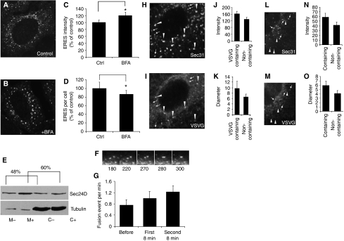
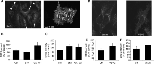
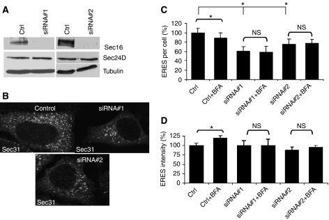
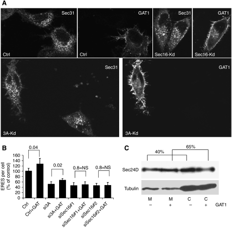
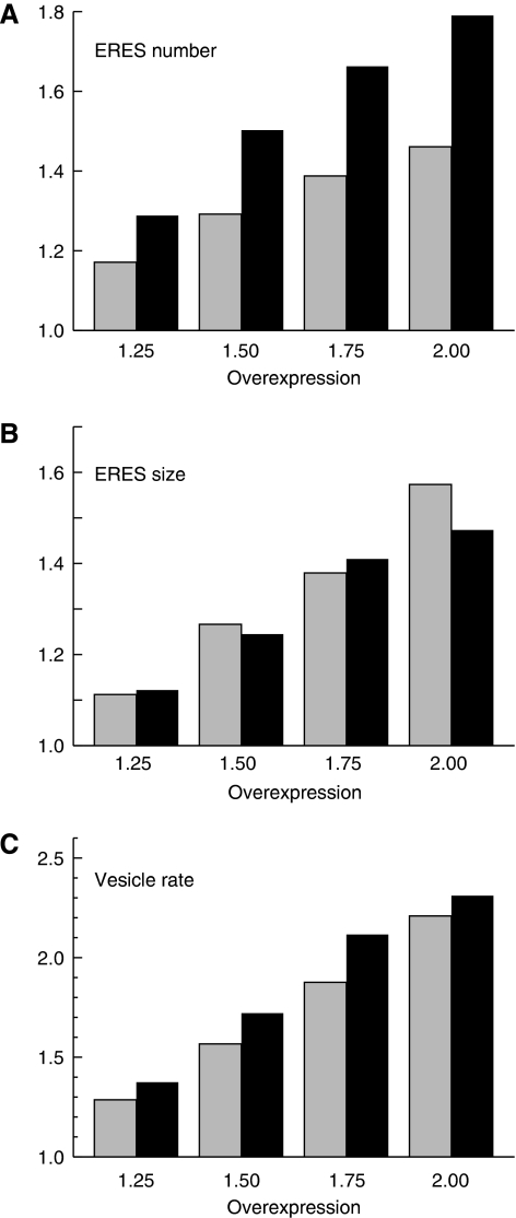
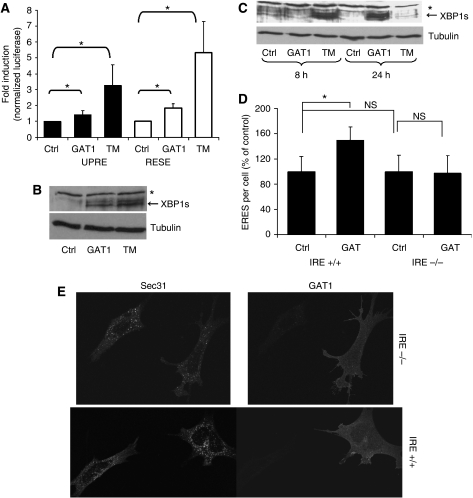
The biogenesis of endoplasmic reticulum (ER) exit sites (ERES) involves the formation of phosphatidylinositol-4 phosphate (PI4) and Sec16, but it is entirely unknown how ERES adapt to variations in cargo load. Here, we studied acute and chronic adaptive responses of ERES to an increase in cargo load for ER export. The acute response (within minutes) to increased cargo load stimulated ERES fusion events, leading to larger but less ERES. Silencing either PI4-kinase IIIalpha (PI4K-IIIalpha) or Sec16 inhibited the acute response. Overexpression of secretory cargo for 24 h induced the unfolded protein response (UPR), upregulated COPII, and the cells formed more ERES. This chronic response was insensitive to silencing PI4K-IIIalpha, but was abrogated by silencing Sec16. The UPR was required as the chronic response was absent in cells lacking inositol-requiring protein 1. Mathematical model simulations further support the notion that increasing ERES number together with COPII levels is an efficient way to enhance the secretory flux. These results indicate that chronic and acute increases in cargo load are handled differentially by ERES and are regulated by different factors.


Figures

Figure 1
Figure 1
Acute cargo overload enlarges ERES and induces ERES fusion activity. (AD) HeLa cells were treated with a solvent (A) or with 5 μg/ml BFA (B) for 40 min at 37°C and ERES were visualized by confocal immunofluorescence microscopy with an antibody against Sec31. Image intensity (C) and number (D) of ERES were evaluated. The data are displayed as a percentage of control to normalize for inter-assay variance. Asterisks indicate statistically significant differences (P<0.05). (E) HeLa cells were treated with a solvent (−) or 5 μg/ml BFA (+) for 40 min. Microsomes (M) and cytosol (C) were prepared and 8 μg of each sample was separated by SDS–PAGE. Sec24D and tubulin were detected by immunoblotting. Sec24D was quantified by densitometry and displayed as percent membrane-bound of total. (F, G) HeLa cells were transfected with a plasmid encoding YFP-tagged Sec23A. Live cell imaging was performed and fusion events were counted. A typical fusion event is shown in (E). Fusion events were counted in a period of 8 min prior (before, n=11) and after the addition (first 8 min, n=11) of 5 μg/ml of BFA. In some cases (n=4), fusion events were also scored for a second period of 8 min (second 8 min) after BFA addition. (HK) HeLa cells were transfected with a plasmid encoding GFP-tagged VSVG-tsO45 and cultured overnight at 40°C. The cells were shifted to 32°C for 5 min and immunostained for Sec31. Images were acquired for Sec31 (H) and for GFP-tagged VSVG-tsO45 (I), and the intensity (J) and diameter (K) of ERES were determined. (LO) HeLa cells were transfected with a plasmid encoding GFP-tagged VSVG-tsO45 and cultured overnight at 40°C. The cells were shifted to 10°C for 40 min and immunostained for Sec31. Images were acquired for Sec31 (L) and for GFP-tagged VSVG-tsO45 (M), and the intensity (N) and diameter (O) of ERES were determined.
Figure 2
Figure 2
Overexpression of cargo proteins increases the size and number of ERES. (A) HeLa cells were transfected with a plasmid encoding YFP-tagged wild-type GAT1. After 24 h, the cells were stained for Sec31, and images were acquired by confocal microscopy. Arrows, transfected cells; arrowheads, non-transfected cells. Graphic representation of the number (B) and intensity (C) of ERES of control (ctrl) cells, cells treated with 5 μg/ml BFA for 40 min (BFA) or cells overexpressing wild-type GAT1 (GAT-WT). (D) HeLa cells were transfected with plasmids encoding GFP-tagged VSVG-tsO45 and cultured at the permissive temperature. After 24 h, the cells were fixed, stained for Sec31, and analysed by confocal microscopy. (E, F) Graphic representation of the number (E) and intensity (F) of ERES of control (ctrl) cells or cells overexpressing VSVG-tsO45 (VSVG).
Figure 3
Figure 3
Effect of knockdown of PI4K-IIIα on ERES and their response to BFA. (A) HeLa cells were transfected with a control siRNA (ctrl) and fixed 72 h later, or with an siRNA to PI4K-IIIα and fixed 72 h later (si3A), or transfected after 48 h with a plasmid encoding HA-tagged PI4K-IIIα and fixed 24 h later (rescue). Fixed cells were stained for Sec31 to label ERES. Evaluation of the number of ERES per cell (B) and their average intensity (C). The results are presented as a percentage of control to account for inter-assay variance. HeLa cells were transfected with a control siRNA. After 72 h, they were treated with a solvent (ctrl) or with 5 μg/ml BFA (ctrl+BFA). HeLa cells were transfected with an siRNA to PI4K-IIIα (si3A). After 72 h, they were treated with a solvent (si3A) or with 5 μg/ml BFA (si3A+BFA). HeLa cells were transfected with an siRNA to PI4K-IIIα. After 48 h, they were transfected with a plasmid encoding HA-tagged PI4K-IIIα and 24 h later they were treated with a solvent (rescue) or with 5 μg/ml BFA (rescue+BFA). (D) HeLa cells were transfected with an siRNA to PI4K-IIIα (+) or with a control siRNA (−). After 72 h, microsomes (M) and cytosol (C) were prepared (see ‘Materials and methods') and 8 μg of each sample was separated by SDS–PAGE. Sec24D, PI4K-IIIα and tubulin were detected by immunoblotting. Sec24D was quantified by densitometry and displayed as percent membrane-bound of total. (E) HeLa cells were transfected with a control siRNA (ctrl) or with an siRNA against PI4K-IIIα (Kd). After 48 h, the cells were transfected with a plasmid encoding YFP-tagged Sec23A as a marker for ERES. Live cell imaging was performed 24 h later. Fusion events were monitored during a period of 8 min before (-before) and 8 min after (-after) addition of 5 μg/ml BFA. *Statistically significant differences (P⩽0.01; non-paired Student's t-test); NS, nonsignificant differences.
Figure 4
Figure 4
Effect of knockdown of Sec16 on ERES. (A) HeLa cells were transfected with a control siRNA (ctrl) or two different siRNAs against Sec16 (siRNA#1, siRNA#2). After 72 h, the cells were lysed and subjected to SDS–PAGE followed by immunoblotting with antibodies against Sec16, Sec24D, and tubulin. (B) HeLa cells were transfected with a control siRNA (control) or with siRNAs against Sec16. After 72 h, the cells were fixed and immunostained for Sec31 to label ERES. (C) Number of ERES per cell and their average intensity (D). *Statistically significant differences (P⩽0.01, non-paired Student's t-test); NS, nonsignificant differences.
Figure 5
Figure 5
Effect of PI4K-IIIα or Sec16 knockdown on the adaptation of ERES to GAT1 overexpression. HeLa cells were transfected with a control siRNA (ctrl), siRNA to PI4K-IIIα (3A-Kd) or siRNA to Sec16 (Sec16-Kd). After 48 h, the cells were either not transfected or transfected with a plasmid encoding YFP-tagged GAT1 (+GAT) and after another 24 h processed for immunofluorescence to label ERES by anti-Sec31. (A) Representative images. (B) Graphical representation of the number of ERES per cell expressed as a percentage of control. Numbers above the bars indicate P-values (paired Student's t-test). (C) HeLa cells grown in six-well plates were transfected 24 h after plating with a plasmid encoding YFP-tagged GAT1 (+) or with an empty plasmid (−). After 24 h, microsomes (M) and cytosol (C) were prepared and 8 μg protein of each sample was separated by SDS–PAGE. Sec24D and tubulin (loading control) were detected by immunoblotting. Membrane-bound Sec24 is indicated as a percentage of total. Note that GAT1 overexpression increases the membrane-bound fraction of Sec24.
Figure 6
Figure 6
Effect of GAT1 overexpression on Sec24D and Sec16. (A) HeLa cells were transfected with a plasmid encoding YFP-tagged GAT1 (+) or with an empty plasmid (−). After 24 h, total cell lysates were separated by SDS–PAGE. Sec24D and Sec16 were detected by immunoblotting and quantified using tubulin as a loading control. Numbers at the right margin of the immunoblots indicate the percentage of induction of expression in the sample expressing GAT1 versus the sample transfected with the empty plasmid. (B) HeLa cells were transfected with siRNA#1 against Sec16. After 24 h, the medium was removed and the cells were either transfected with a plasmid encoding YFP-tagged GAT1 (+) or with an empty plasmid (−). The cells were lysed after 24 h and 8 μg of lysate was separated by SDS–PAGE. Sec16 was detected by immunoblotting and quantified using tubulin as a loading control. The upper panel shows an outline of the experiment. Numbers at the right margin of the immunoblot indicate the percentage of induction of Sec16 by GAT1 overexpression.
Figure 7
Figure 7
Simulation of the effect of increasing COPII levels on ERES size and number, and on the rate of COPII vesicle production. Increase in the mean number (A) and size (B) of ERES with rising cellular COPII levels. The amount of overexpression is determined with regard to the putative native level investigated in Heinzer et al (2008). Grey bars represent the data for a slow COPII turnover (kon=koff=0.1/s), whereas black bars indicate the respective values for a faster turnover (kon=koff=0.2/s). The increase in ERES size and number, as a function of COPII levels, is very similar for both turnover rates. (C) The rate of vesicle production continues to increase proportionally with increasing COPII levels for both turnover rates (grey bars: slow turnover; black bars: fast turnover).
Figure 8
Figure 8
Chronic cargo overload triggers the UPR. (A) HeLa cells grown in 12-well plates were transfected with expression vectors encoding firefly luciferase under the control of ERSE or UPRE. Transfections included an empty plasmid (ctrl) or YFP-tagged GAT1 (GAT1). Renilla luciferase was used to normalize for transfection efficiency. The ratio of plasmid to ERSE/UPRE to Renilla was always 2:2:1. The luciferase assay was performed 24 h after transfection. TM, treatment with tunicamycin (10 μg/ml) for 5 h. (B) HeLa cells grown in six-well plates were transfected with an empty plasmid (ctrl) or with a plasmid encoding YFP-tagged GAT1 (GAT1). On the following day, one well transfected with the empty plasmid was treated with tunicamycin (10 μg/ml) for 8 h (TM). The cells were lysed after 24 h in RIPA buffer and separated by SDS–PAGE. XBP1s was detected by immunoblotting. *Nonspecific band. The immunoblot was stripped and tubulin was stained to demonstrate equal loading. (C) HeLa cells grown in six-well plates were transfected with an empty plasmid or with a plasmid encoding YFP-tagged GAT1 (GAT1). Two samples transfected with an empty plasmid were treated either with solvent (ctrl) or with 10 μg/ml tunicamycin (TM) for 8 and 24 h. The cells were lysed after 8 or 24 h in RIPA buffer and separated by SDS–PAGE. XBP1s was detected by immunoblotting. *Nonspecific band. The stripped blot was re-stained for tubulin to demonstrate equal loading. (D) MEFs derived from wild-type mice (IRE+/+) or mice lacking IRE1 (IRE−/−) were electroporated with a plasmid encoding YFP-tagged GAT1. After 24 h, the cells were fixed and ERES were stained with anti-Sec31. (C) A graphic representation of the number of ERES per cells as a comparison between cells that express GAT1 (GAT1) and cells that do not (ctrl). Cells of comparable size were chosen for this analysis. (E) Representative images of ERES in cells expressing or not expressing GAT1. *Statistically significant differences (P⩽0.05; paired Student's t-test); NS, nonsignificant differences.

References

    1. Acosta-Alvear D, Zhou Y, Blais A, Tsikitis M, Lents NH, Arias C, Lennon CJ, Kluger Y, Dynlacht BD (2007) XBP1 controls diverse cell type- and condition-specific transcriptional regulatory networks. Mol Cell 27: 53–66 - PubMed
    1. Bevis BJ, Hammond AT, Reinke CA, Glick BS (2002) De novo formation of transitional ER sites and Golgi structures in Pichia pastoris. Nat Cell Biol 4: 750–756 - PubMed
    1. Bhattacharyya D, Glick BS (2007) Two mammalian Sec16 homologues have nonredundant functions in endoplasmic reticulum (ER) export and transitional ER organization. Mol Biol Cell 18: 839–849 - PMC - PubMed
    1. Bi X, Corpina RA, Goldberg J (2002) Structure of the Sec23/24-Sar1 pre-budding complex of the COPII vesicle coat. Nature 419: 271–277 - PubMed
    1. Blumental-Perry A, Haney CJ, Weixel KM, Watkins SC, Weisz OA, Aridor M (2006) Phosphatidylinositol 4-phosphate formation at ER exit sites regulates ER export. Dev Cell 11: 671–682 - PubMed

Publication types

MeSH terms

Substances