. 2008 Aug 20;27(16):2250-8.
doi: 10.1038/emboj.2008.135. Epub 2008 Jul 24.

Cryo-electron microscopy reveals a novel DNA-binding site on the MCM helicase

Affiliations

Cryo-electron microscopy reveals a novel DNA-binding site on the MCM helicase

Alessandro Costa et al. EMBO J. .

Abstract

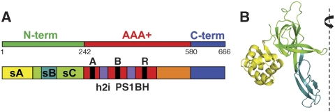
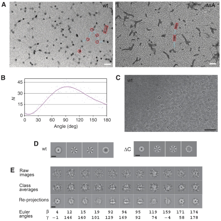
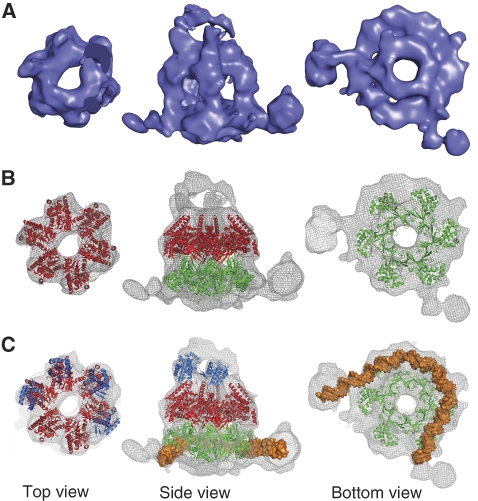
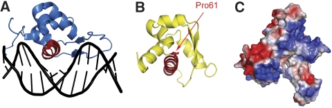
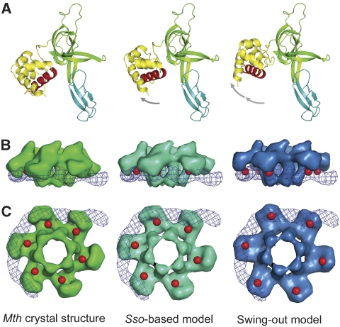
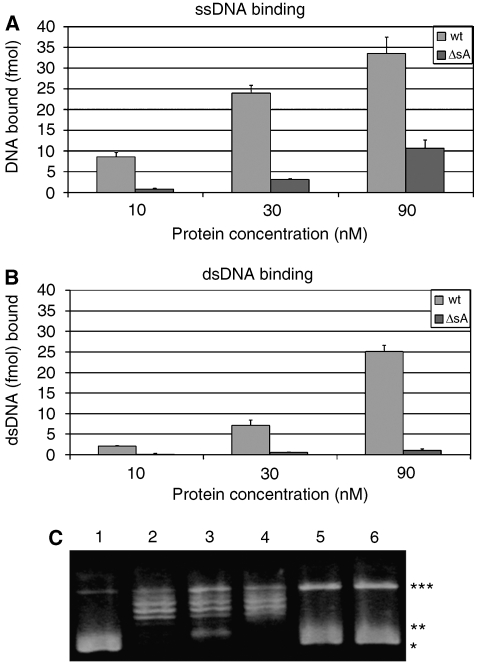
The eukaryotic MCM2-7 complex is recruited at origins of replication during the G1 phase and acts as the main helicase at the replication fork during the S phase of the cell cycle. To characterize the interplay between the MCM helicase and DNA prior to the melting of the double helix, we determined the structure of an archaeal MCM orthologue bound to a 5.6-kb double-stranded DNA segment, using cryo-electron microscopy. DNA wraps around the N-terminal face of a single hexameric ring. This interaction requires a conformational change within the outer belt of the MCM N-terminal domain, exposing a previously unrecognized helix-turn-helix DNA-binding motif. Our findings provide novel insights into the role of the MCM complex during the initiation step of DNA replication.


Figures

Figure 1
Figure 1
Domain structure of MthMCM. (A) Schematic representation of the primary structure of MthMCM, comprising a N-terminal domain (green) a AAA+ domain (red) and a C-terminal domain (blue). The N-terminal domain can be further subdivided into 3 subdomains (sA, sB, sC, in yellow, teal and green, respectively). The AAA+ domain folds into an α/β-subdomain (red) and a α-domain (orange); the former contains the canonical Walker A, Walker B and R finger motifs found in AAA+ protein, as well as the unique insertions (h2i and PS1BH, in violet), which characterize the MCM clade. The C-terminal domain is predicted to fold into a winged-helix motif. (B) The crystal structure of a monomer of the MthMCM N-terminal domain (Fletcher et al, 2003; PDB entry 1LTL), with subdomains sA, sB and sC colour-coded as in (A). A dashed line indicates the position of the six-fold axis around which the hexameric complex assembles.
Figure 2
Figure 2
Electron microscopy of MthMCM bound to long dsDNA molecules. (A) Micrographs of a positively stained complex between dsDNA (5600 bp) and either wt MthMCM (left panel) or the protein lacking subdomain A (ΔsA, right panel). Images were taken at a magnification of × 27 500 and 5 μm defocus. A few features have been highlighted: MCM complexes are shown as red rings, rectangular boxes indicate the presence of fibres, whereas DNA segments are highlighted in cyan. Scale bar 500 Å. (B) Statistical analysis of angle distribution performed on a sample of 5000 protein-induced dsDNA kinks carried out on the wt MthMCM-negative stain data. (C) Micrograph of MthMCM bound to long dsDNA molecules embedded in vitreous ice. Scale bar 500 Å. (D) Eigenimages obtained from multivariate statistical analysis performed on translationally aligned cryo-EM particles, both for the wt (left panel) and a mutant lacking the C-terminal domain (ΔC, right panel). Scale bar 100 Å. (E) Overview of the 3D reconstruction procedure. Scale bar 100 Å.
Figure 3
Figure 3
Cryo-EM structure of wt MthMCM bound to long dsDNA molecules. (A) Surface rendering of the molecule (displayed at 2.5 σ) showing a slab through the top, side and bottom views, respectively. The structure consists of a single ring with a large central cavity. Whereas the top face of the ring displays six-fold symmetry, the bottom face is rounded, with two filamentous protrusions departing radially. (B) Fitting of the AAA+ domain from MkaMCM (red; Bae et al, submitted) and the N-terminal domain from MthMCM (green; Fletcher et al, 2003). The AAA+ hexamer fits well to the dome-shaped top face, whereas additional electron density surrounds the atomic model in the bottom tier. (C) Four copies of the WH domain of Cdc6 (blue; Liu et al, 2000) have been fitted to the protrusions from the AAA+ domain (Supplementary Figure 4). Roughly 80 bp of dsDNA has been fitted to the additional electron density at the bottom of the ring (Supplementary Figure 5); a re-orientation of subdomain A is required to avoid steric clashes with the modelled dsDNA.
Figure 4
Figure 4
Subdomain A contains a helix-turn-helix. (A) The crystal structure of the Genesis transcription factor bound to dsDNA (Jin et al, 1999; PDB entry 2HDC) with the recognition helix is highlighted in red. (B) The structure of subdomain A in the same orientation: the atomic model of proline 61 is shown in red, respectively. (C) Surface electrostatic potential of the N-terminal domain, in the same orientation as in Figure 1B, with negative charges shown in red and positive charges in blue.
Figure 5
Figure 5
Modelling of the swing-out movement of subdomain A. The left panel shows the crystal structure of the N-terminal domain of MthMCM (Fletcher et al, 2003), the central panel the same structure with subdomain A remodelled based on the crystal structure of the S. solfataricus MCM N-terminal domain (Liu et al, 2008), and the right panel the structure modelled to fit the EM data. The modelling was carried out by extrapolating the movement derived from the differences between the MthMCM and SsoMCM structures. (A) A ribbon diagram representation of the N-terminal domain monomer, colour-coded as in Figure 1B, with the helix beginning with proline 61 highlighted in red. The left panel shows that the putative recognition helix packs against subdomain C, whereas in the conformation shown on the right panel is available for interaction with dsDNA. (B) Side view surface representation of the hexameric N-terminal domain obtained by filtering to 20-Å resolution the electron density calculated from the models shown above, overlaid with a surface representation of the modelled dsDNA (depicted as a blue mesh). The location of the recognition helix is highlighted in red. (C) The same surface representation viewed from the bottom.
Figure 6
Figure 6
Biochemical characterization of the MthMCM–DNA interaction. DNA-binding properties of the ΔsA mutant: filter-binding assays were performed using both the wt and ΔsA mutant proteins, in the presence of (A) a ssDNA substrate of 50 nucleotides and (B) a 50 bp dsDNA substrate. The ΔsA mutant shows a decreased binding to both ssDNA and dsDNA. Whereas the binding of ssDNA increases with the amount of protein, binding to short stretches of dsDNA (50 bp) remains virtually undetectable. (C) Topology footprint assay. Plasmid DNA relaxation at saturating concentration of Topoisomerase I and in the presence of MthMCM demonstrates a role of the MCM complex in the modulation of plasmid topological state and confirms the involvement of subdomain A. Lane 1: 5.7 nM pUC18. Lane 2: pUC18 incubated with 20 U of wheat germ Topoisomerase I. Lane 3: pUC18 incubated with 1 μM of wt MthMCM (hexamer), for 15 min, followed by a 60 min incubation with Topoisomerase I. Lane 4: the same experiment as in lane 3, where the ΔsA mutant is used instead of wt MthMCM. Lanes 5 and 6: pUC18 incubated with 1 μM of MthMCM or ΔsA, respectively. Different topological states can be visualized: *negatively supercoiled DNA; **partially relaxed DNA; ***totally relaxed DNA.

Similar articles

Cited by

References

    1. Aravind L, Anantharaman V, Balaji S, Babu MM, Iyer LM (2005) The many faces of the helix-turn-helix domain: transcription regulation and beyond. FEMS Microbiol Rev 29: 231–262 - PubMed
    1. Arias EE, Walter JC (2007) Strength in numbers: preventing rereplication via multiple mechanisms in eukaryotic cells. Genes Dev 21: 497–518 - PubMed
    1. Barry ER, McGeoch AT, Kelman Z, Bell SD (2007) Archaeal MCM has separable processivity, substrate choice and helicase domains. Nucleic Acids Res 35: 988–998 - PMC - PubMed
    1. Boekema EJ, Berden JA, van Heel MG (1986) Structure of mitochondrial F1-ATPase studied by electron microscopy and image processing. Biochim Biophys Acta 851: 353–360 - PubMed
    1. Boisset N, Penczek PA, Taveay JC, You V, de Haas F, Lamy J (1998) Overabundant single-particle electron microscope views induce a three-dimensional reconstruction artifact. Ultramicroscopy 74: 201–207

Publication types