. 2008 Aug;237(8):2209-19.
doi: 10.1002/dvdy.21634.

Isolation and expression of Pax6 and atonal homologues in the American horseshoe crab, Limulus polyphemus

Affiliations

Isolation and expression of Pax6 and atonal homologues in the American horseshoe crab, Limulus polyphemus

David C Blackburn et al. Dev Dyn. 2008 Aug.

Abstract

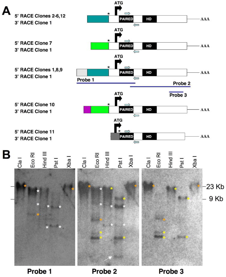
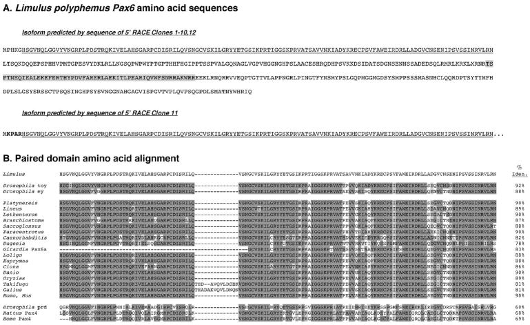
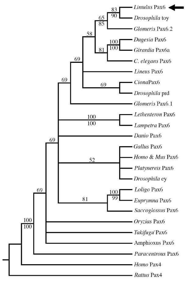
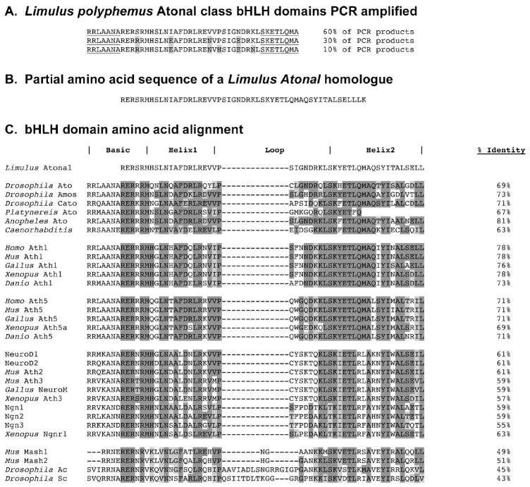
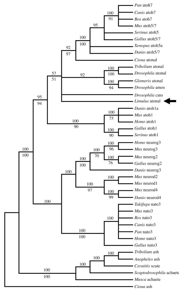
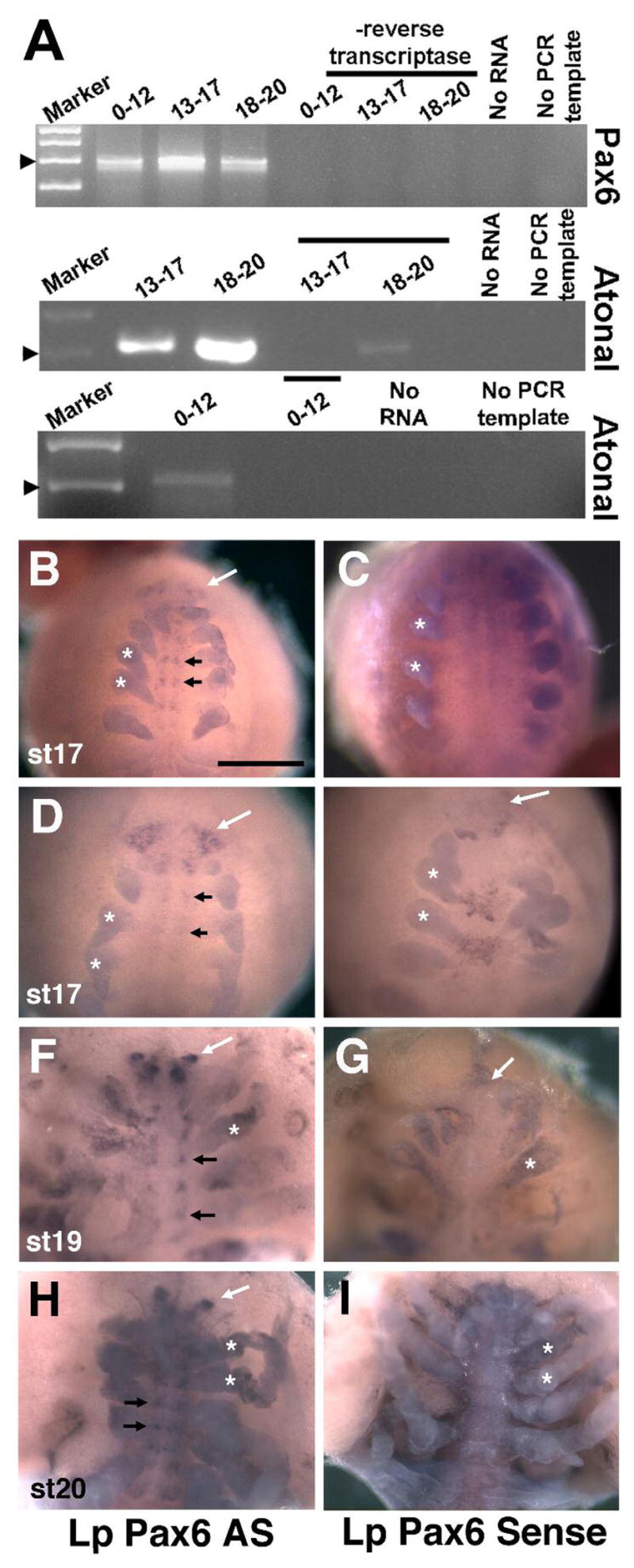
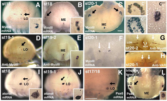
Pax6 regulates eye development in many animals. In addition, Pax6 activates atonal transcription factors in both invertebrate and vertebrate eyes. Here, we investigate the roles of Pax6 and atonal during embryonic development of Limulus polyphemus rudimentary lateral, medial and ventral eyes, and the initiation of lateral ommatidial eye and medial ocelli formation. Limulus eye development is of particular interest because these animals hold a unique position in arthropod phylogeny and possess multiple eye types. Furthermore, the molecular underpinnings of eye development have yet to be investigated in chelicerates. We characterized a Limulus Pax6 gene, with multiple splice products and predicted protein isoforms, and one atonal homologue. Unexpectedly, neither gene is expressed in the developing eye types examined, although both genes are present in the lateral sense organ, a structure of unknown function.


Figures

Figure 1
Figure 1. Lp Pax6 encodes multiple isoforms
A) Diagram of five different types Lp Pax6 5′ RACE products with a single 3′ RACE product. Color-coding indicates different 5′ UTR sequences. Use of the same color among different classes of clones indicates stretches of exact nucleotide identity. The positions of gene-specific RACE primers are indicated by blue arrows, below (5′ RACE) or above (3′ RACE) each diagram. 11 of 12 5′ RACE products have identical sequences beginning at a putative splice acceptor site (asterisk) upstream of the start ATG codon. One 5′ RACE subclone (#11) has an alternative in frame ATG start, plus 4 unique amino acids immediately upstream of the paired domain (bold-faced amino acids in Figure 2A). The longest 5′ RACE product (probe 1) was used for Southern blot analyses. B) Autoradiographs of replicate Limulus genomic Southern blots hybridized with the cDNA probes shown in A. Enzymes used to digest Limulus genomic DNA are indicated above each lane. White asterisks to the right of bands indicate those in common for Probes 1 and 2, yellow asterisks mark bands in common between Probes 2 and 3, and orange asterisks indicate bands common to all three probes. Several of the bands marked by white asterisks gave a weaker signal on the Southern blot autoradiogram. The white arrow in the middle blot points to the only band that uniquely hybridizes with Probe 2.
Figure 2
Figure 2. Amino acid sequence of Lp Pax6
(A) Deduced amino acid sequence of two predicted protein isoforms of Limulus Pax6 that only differ by four amino acids (bold-faced in clone 11). The paired domain is underlined and the homeodomain shaded. (B) Amino acid sequence comparison of the paired domain for Lp Pax6 with various invertebrate and vertebrate Pax6 proteins in the NIH/NCBI database. Dark shading indicates identical amino acids, light shading similar amino acids to Lp Pax6. Percent identities (uncorrected pairwise sequence divergences) are listed at the right. The Lp Pax6 paired domain shares the highest percent identity with that of Drosophila toy.
Figure 3
Figure 3. Phylogenetic analyses of Lp Pax6
Majority rule consensus tree of the 114805 most parsimonious trees resulting from a heuristic search using maximum parsimony; numbers above branches are the percentage of trees in which the topology appears. Numbers below branches are the posterior probabilities for that topology found through a Bayesian phylogenetic analysis using a mixed amino acid model. Pax6 paired domains form a clade that is strongly supported by both analyses. Among the Pax6 paired domains, there is strong support that the Lp Pax6 paired domain (black arrow) is most similar to the Drosophila toy gene. There is also strong support from both analyses for clades comprising the paired domains of planarian flatworms (Dugesia and Girardia), lampreys (Lethenteron and Lampetra) and mollusks (Loligo and Euprymna).
Figure 4
Figure 4. Molecular characterization of Lp atonal
A) Amino acid sequences of three types of atonal-related bHLH domains PCR amplified from Limulus genomic DNA. Degenerate PCR primers were designed for the underlined amino acids, based on Drosophila atonal and Xenopus Ath5 bHLH domains (Brown et al., 1998). B) Partial amino acid sequence of a Limulus atonal homologue partially cloned. C) Comparison of bHLH domain amino acid sequence for Lp atonal with those of various invertebrate and vertebrate Atonal/Ath/ATOH proteins in the NIH/NCBI database. Dark shading indicates identical amino acids, light shading similar amino acids to Lp Atonal. Percent identities (uncorrected pairwise sequence divergences) are listed in the right column. The Limulus bHLH domain shares the highest percent identity with the Anopheles (mosquito) atonal gene.
Figure 5
Figure 5. Phylogenetic analyses of Lp Atonal
Majority rule consensus tree of the 37 most parsimonious trees found in a heuristic search using maximum parsimony; numbers above branches are the percentage of the trees in which the topology appears. Numbers below branches are the posterior probabilities for that topology found through a Bayesian phylogenetic analysis, using a mixed amino acid model. Ath5/ATOH7, Ath1/ATOH1, and atonal bHLH domains form a clade that is strongly supported by both analyses. Among these bHLH domains, the relationship of the Lp atonal bHLH domain (black arrow) to those of other atonal genes is unresolved, although there is weak support that it may be more closely related to vertebrate Ath5 and atonal genes.
Figure 6
Figure 6. Embryonic expression of Limulus Pax6 and Atonal mRNA
A) RT-PCR for Lp Pax6 and Lp Atonal indicating both genes are expressed throughout embryogenesis to hatching at late st 20. All PCR and RT controls exhibited little to no background amplification. B–I) In situ hybridization for Lp Pax6 antisense (AS) and sense probes, with all panels showing ventral views. B–E) St 17 embryos with Lp Pax6 mRNA expression in the forming brain (white arrows) and ventral nerve cord (black arrows), compared to sense controls. F–G) St 19 embryo with discrete brain (white arrow) and clustered ventral nerve cord (black arrows) expression domains. H,I) St 20 embryo with brain lobe (white arrows) and clustered ventral nerve cord expression (black arrows). Asterisks in all panels label forming appendages that are nonspecifically stained in both antisense and sense probed embryos. Embryo staging followed Harzsch et al, 2006. Bar in B = 500μm.
Figure 7
Figure 7. Limulus Pax6 and atonal are not expressed in developing eyes
A–C,F) Myosin III mRNA expression in the rudimentary lateral eye (black arrows in A–C), early lateral ommatidial eyes (C″, C‴ show first ommatidial photoreceptors surrounded by crescent of rudimentary photoreceptors), rudimentary median eye (ME; B, C, C‴) and rudimentary ventral photoreceptors (arrows in F). The lateral sense organ (LO) is not involved in vision and does not express MyoIII mRNA (A and data not shown). D,E) Anti-Myo III antibody labels the embryonic rudimentary lateral eye (arrow in D), median eye (E) and more weakly the LO (D). F) High magnification of ventral photoreceptors (arrows) expressing MyoIII mRNA. G–H) Anti-MyoIII (G) and Anti-Visual Arrestin (VAR) staining of ventral photoreceptors (arrows) at approximately the same age as in F. These antibodies also label forming optic nerves innervating the brain from the rudimentary lateral (LON) and median (MON) eyes. I–J). Lp Atonal expression in the LO but not rudimentary lateral eye (black arrows point to eye). Insets show higher magnification of LO. K–L) Throughout ages when MyoIII mRNA and protein are expressed in rudimentary eyes or forming lateral ommatidial eyes, Pax6 mRNA is observed in the LO but is absent from these eyes (arrow points to rudimentary lateral eye). Panels A,D,I,J and K are lateral, B and E frontal, C and L are top-down dorsal, and F–H are ventral views. Scale bars in A, C = 500 μm, inset magnification is an additional 400X.

Similar articles

Cited by

References

    1. Abzhanov A, Popadic A, Kaufman TC. Chelicerate Hox genes and the homology of arthropod segments. Evol Dev. 1999;1:77–89. - PubMed
    1. Arendt D. Evolution of eyes and photoreceptor cell types. Int J Dev Biol. 2003;47:563–571. - PubMed
    1. Arendt D, Tessmar K, de Campos-Baptista MI, Dorresteijn A, Wittbrodt J. Development of pigment-cup eyes in the polychaete Platynereis dumerilii and evolutionary conservation of larval eyes in Bilateria. Development. 2002;129:1143–1154. - PubMed
    1. Arendt D, Wittbrodt J. Reconstructing the eyes of Urbilateria. Philos Trans R Soc Lond B Biol Sci. 2001;356:1545–1563. - PMC - PubMed
    1. Avise JC, Nelson WS, Sugita H. A special history of “living fossils”: molecular evolutionary patterns in horseshoe crabs. Evolution. 1994;48:1986–2001. - PubMed

Publication types

MeSH terms

LinkOut - more resources