

Structure and mechanistic implications of a uroporphyrinogen III synthase-product complex
- PMID: 18651750
- PMCID: PMC2852885
- DOI: 10.1021/bi800635y
Structure and mechanistic implications of a uroporphyrinogen III synthase-product complex
Abstract
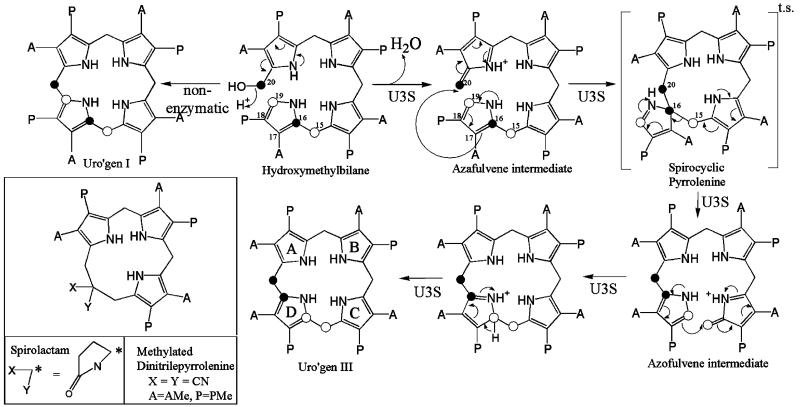
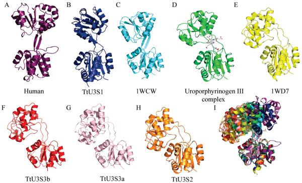
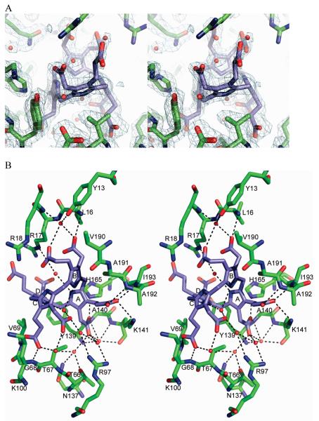
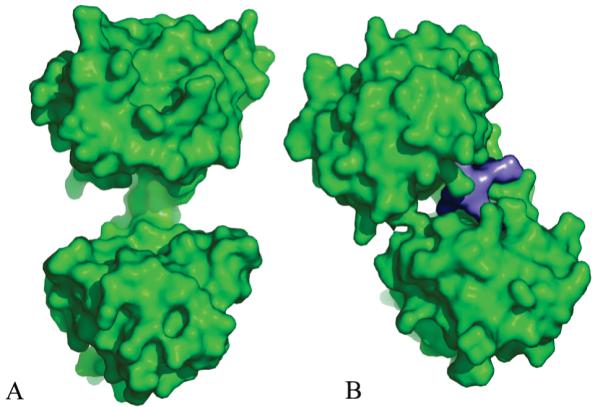
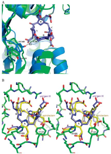
Uroporphyrinogen III synthase (U3S) catalyzes the asymmetrical cyclization of a linear tetrapyrrole to form the physiologically relevant uroporphyrinogen III (uro'gen III) isomer during heme biosynthesis. Here, we report four apoenzyme and one product complex crystal structures of the Thermus thermophilus (HB27) U3S protein. The overlay of eight crystallographically unique U3S molecules reveals a huge range of conformational flexibility, including a "closed" product complex. The product, uro'gen III, binds between the two domains and is held in place by a network of hydrogen bonds between the product's side chain carboxylates and the protein's main chain amides. Interactions of the product A and B ring carboxylate side chains with both structural domains of U3S appear to dictate the relative orientation of the domains in the closed enzyme conformation and likely remain intact during catalysis. The product C and D rings are less constrained in the structure, consistent with the conformational changes required for the catalytic cyclization with inversion of D ring orientation. A conserved tyrosine residue is potentially positioned to facilitate loss of a hydroxyl from the substrate to initiate the catalytic reaction.
Figures
Similar articles
-
Crystal structure of uroporphyrinogen III synthase from Pseudomonas syringae pv. tomato DC3000.Biochem Biophys Res Commun. 2011 May 20;408(4):576-81. doi: 10.1016/j.bbrc.2011.04.064. Epub 2011 Apr 19. Biochem Biophys Res Commun. 2011. PMID: 21527255
-
Human uroporphyrinogen III synthase: NMR-based mapping of the active site.Proteins. 2008 May 1;71(2):855-73. doi: 10.1002/prot.21755. Proteins. 2008. PMID: 18004775
-
Crystal structure of human uroporphyrinogen III synthase.EMBO J. 2001 Nov 1;20(21):5832-9. doi: 10.1093/emboj/20.21.5832. EMBO J. 2001. PMID: 11689424 Free PMC article.
-
Structural diversity in metal ion chelation and the structure of uroporphyrinogen III synthase.Biochem Soc Trans. 2002 Aug;30(4):595-600. doi: 10.1042/bst0300595. Biochem Soc Trans. 2002. PMID: 12196144 Review.
-
Porphobilinogen deaminase and uroporphyrinogen III synthase: structure, molecular biology, and mechanism.J Bioenerg Biomembr. 1995 Apr;27(2):181-95. doi: 10.1007/BF02110033. J Bioenerg Biomembr. 1995. PMID: 7592565 Review.
Cited by
-
The AEROPATH project targeting Pseudomonas aeruginosa: crystallographic studies for assessment of potential targets in early-stage drug discovery.Acta Crystallogr Sect F Struct Biol Cryst Commun. 2013 Jan 1;69(Pt 1):25-34. doi: 10.1107/S1744309112044739. Epub 2012 Dec 25. Acta Crystallogr Sect F Struct Biol Cryst Commun. 2013. PMID: 23295481 Free PMC article.
-
Crystal structure of the heme d1 biosynthesis enzyme NirE in complex with its substrate reveals new insights into the catalytic mechanism of S-adenosyl-L-methionine-dependent uroporphyrinogen III methyltransferases.J Biol Chem. 2011 Jul 29;286(30):26754-67. doi: 10.1074/jbc.M111.239855. Epub 2011 May 31. J Biol Chem. 2011. PMID: 21632530 Free PMC article.
-
Biosynthesis of the modified tetrapyrroles-the pigments of life.J Biol Chem. 2020 May 15;295(20):6888-6925. doi: 10.1074/jbc.REV120.006194. Epub 2020 Apr 2. J Biol Chem. 2020. PMID: 32241908 Free PMC article. Review.
-
Erythroid heme biosynthesis and its disorders.Cold Spring Harb Perspect Med. 2013 Apr 1;3(4):a011676. doi: 10.1101/cshperspect.a011676. Cold Spring Harb Perspect Med. 2013. PMID: 23471474 Free PMC article. Review.
-
One ring to rule them all: trafficking of heme and heme synthesis intermediates in the metazoans.Biochim Biophys Acta. 2012 Sep;1823(9):1617-32. doi: 10.1016/j.bbamcr.2012.04.009. Epub 2012 May 8. Biochim Biophys Acta. 2012. PMID: 22575458 Free PMC article. Review.
References
-
- Chadwick D, Ackrill K, editors. The Biosynthesis of the Tetrapyrrole Pigments. Vol. 180. John Wiley & Sons; New York: 1994.
-
- Dailey H, editor. Biosynthesis of Heme and Chlorophylls. McGraw-Hill; 1990.
-
- Mathewson J, Corwin A. Biosynthesis of pyrrole pigments: a mechanism for porphybilinogen polymerization. J. Am. Chem. Comm. 1961:1313–1315.
-
- Battersby AR, McDonald E. Biosynthesis of porphyrins and corrins. Philos. Trans. R. Soc. London B. 1976;273:161–180. - PubMed
-
- Battersby AR, Fookes CJR, Matcham GWJ, McDonald E. Biosynthesis of the pigments of life: formation of the macrocycle. Nature. 1980;285:17–21. - PubMed
Publication types
MeSH terms
Substances
Associated data
- Actions
- Actions
- Actions
- Actions
Grants and funding
LinkOut - more resources
Full Text Sources
Molecular Biology Databases
