

Brain structural changes in obstructive sleep apnea
- PMID: 18652092
- PMCID: PMC2491498
Brain structural changes in obstructive sleep apnea
Abstract
Study objectives: Determine whether obstructive sleep apnea (OSA) subjects show indications of axonal injury.
Design: We assessed fiber integrity in OSA and control subjects with diffusion tensor imaging (DTI). We acquired four whole-brain DTI series from each subject. The four series were realigned, and the diffusion tensor calculated at each voxel. Fractional anisotropy (FA), a measure of fiber integrity, was derived from the diffusion tensor, resulting in a whole brain FA "map." The FA maps were spatially normalized, smoothed, and compared using voxel-based statistics to determine differences between OSA and control groups, with age as a covariate (P < 0.05, corrected for multiple comparisons).
Setting: University medical center.
Subjects: We studied 41 patients with untreated OSA (mean age +/- SD: 46.3 +/- 8.9 years; female/male: 7/34) with apnea-hypopnea index 15 to 101 (mean +/- SD: 35.7 +/- 18.1 events/hour), and 69 control subjects (mean age +/- SD: 47.5 +/- 8.79 years; female/male: 25/44).
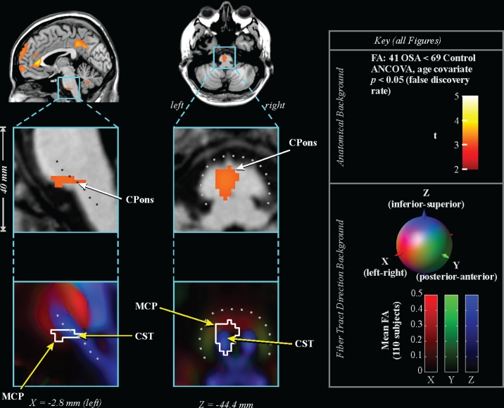
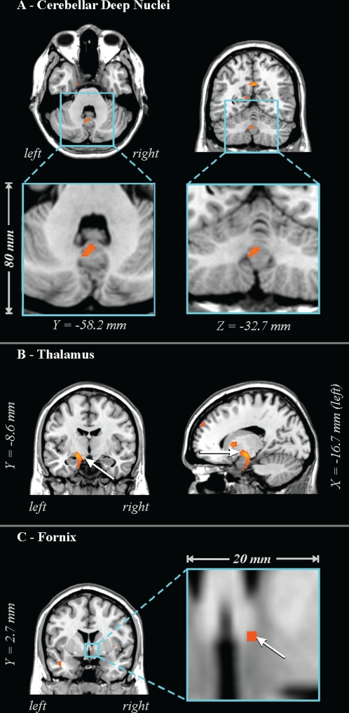
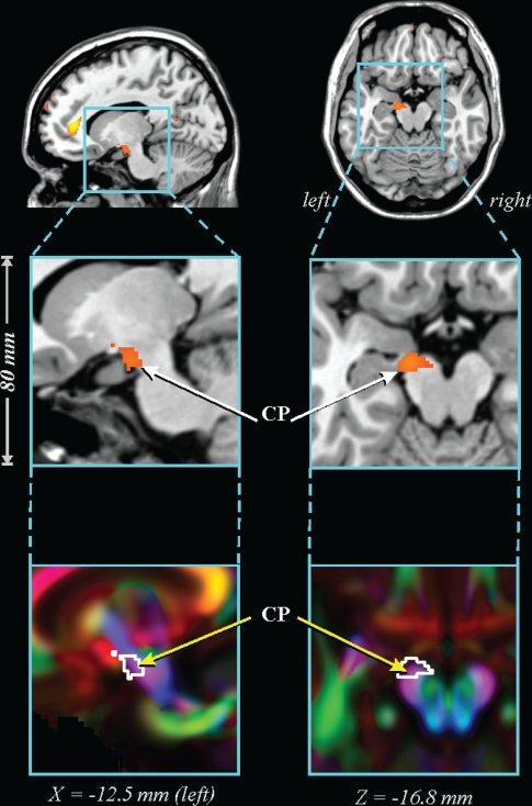
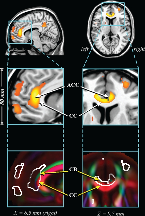
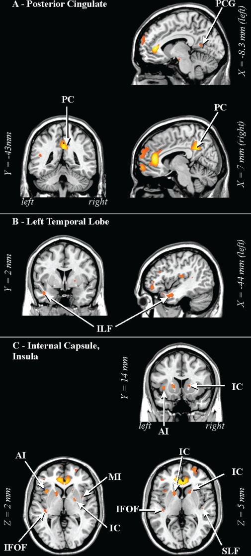
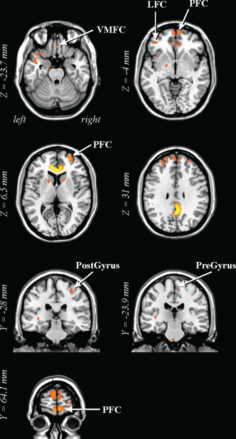
Measurements and results: Multiple regions of lower FA appeared within white matter in the OSA group, and included fibers of the anterior corpus callosum, anterior and posterior cingulate cortex and cingulum bundle, right column of the fornix, portions of the frontal, ventral prefrontal, parietal and insular cortices, bilateral internal capsule, left cerebral peduncle, middle cerebellar peduncle and corticospinal tract, and deep cerebellar nuclei.
Conclusions: White matter is extensively affected in OSA patients; the alterations include axons linking major structures within the limbic system, pons, frontal, temporal and parietal cortices, and projections to and from the cerebellum.
Figures
Similar articles
-
Neural alterations and depressive symptoms in obstructive sleep apnea patients.Sleep. 2008 Aug;31(8):1103-9. Sleep. 2008. PMID: 18714782 Free PMC article.
-
Altered global and regional brain mean diffusivity in patients with obstructive sleep apnea.J Neurosci Res. 2012 Oct;90(10):2043-52. doi: 10.1002/jnr.23083. Epub 2012 Jun 20. J Neurosci Res. 2012. PMID: 22715089 Free PMC article.
-
Sex differences in white matter alterations accompanying obstructive sleep apnea.Sleep. 2012 Dec 1;35(12):1603-13. doi: 10.5665/sleep.2228. Sleep. 2012. PMID: 23204603 Free PMC article.
-
The role of diffusion tensor imaging and fractional anisotropy in the evaluation of patients with idiopathic normal pressure hydrocephalus: a literature review.Neurosurg Focus. 2016 Sep;41(3):E12. doi: 10.3171/2016.6.FOCUS16192. Neurosurg Focus. 2016. PMID: 27581308 Review.
-
White matter alterations in patients with obstructive sleep apnea: a systematic review of diffusion MRI studies.Sleep Med. 2020 Nov;75:236-245. doi: 10.1016/j.sleep.2020.06.024. Epub 2020 Jun 29. Sleep Med. 2020. PMID: 32861061
Cited by
-
Retinal vessel abnormalities as a possible biomarker of brain volume loss in obese adolescents.Obesity (Silver Spring). 2013 Dec;21(12):E577-85. doi: 10.1002/oby.20450. Epub 2013 Jul 2. Obesity (Silver Spring). 2013. PMID: 23512847 Free PMC article.
-
White matter tract-specific alterations in male patients with untreated obstructive sleep apnea are associated with worse cognitive function.Sleep. 2020 Mar 12;43(3):zsz247. doi: 10.1093/sleep/zsz247. Sleep. 2020. PMID: 31848608 Free PMC article.
-
Fatigue in Chronic Respiratory Diseases: Theoretical Framework and Implications For Real-Life Performance and Rehabilitation.Front Physiol. 2018 Sep 19;9:1285. doi: 10.3389/fphys.2018.01285. eCollection 2018. Front Physiol. 2018. PMID: 30283347 Free PMC article. Review.
-
The Impacts of Obstructive Sleep Apnea Severity on Brain White Matter Integrity and Cognitive Functions in Children: A Diffusion Tensor Imaging Study.Nat Sci Sleep. 2021 Dec 2;13:2125-2135. doi: 10.2147/NSS.S329408. eCollection 2021. Nat Sci Sleep. 2021. PMID: 34880696 Free PMC article.
-
Mechanisms of the Rapid Effects of Ketamine on Depression and Sleep Disturbances: A Narrative Review.Front Pharmacol. 2021 Dec 14;12:782457. doi: 10.3389/fphar.2021.782457. eCollection 2021. Front Pharmacol. 2021. PMID: 34970147 Free PMC article. Review.
References
-
- Sateia MJ. Neuropsychological impairment and quality of life in obstructive sleep apnea. Clin Chest Med. 2003;24:249–59. - PubMed
-
- Beebe DW, Groesz L, Wells C, Nichols A, McGee K. The neuropsychological effects of obstructive sleep apnea: a meta-analysis of norm-referenced and case-controlled data. Sleep. 2003;26:298–307. - PubMed
-
- Cortelli P, Parchi P, Sforza E, et al. Cardiovascular autonomic dysfunction in normotensive awake subjects with obstructive sleep apnoea syndrome. Clin Auton Res. 1994;4:57–62. - PubMed
-
- Ferini-Strambi L, Baietto C, Di Gioia MR, et al. Cognitive dysfunction in patients with obstructive sleep apnea (OSA): partial reversibility after continuous positive airway pressure (CPAP) Brain Res Bull. 2003;61:87–92. - PubMed
Publication types
MeSH terms
Grants and funding
LinkOut - more resources
Full Text Sources
Medical