

The N-terminus of apolipoprotein A-V adopts a helix bundle molecular architecture
- PMID: 18652480
- PMCID: PMC2893590
- DOI: 10.1021/bi800515c
The N-terminus of apolipoprotein A-V adopts a helix bundle molecular architecture
Abstract
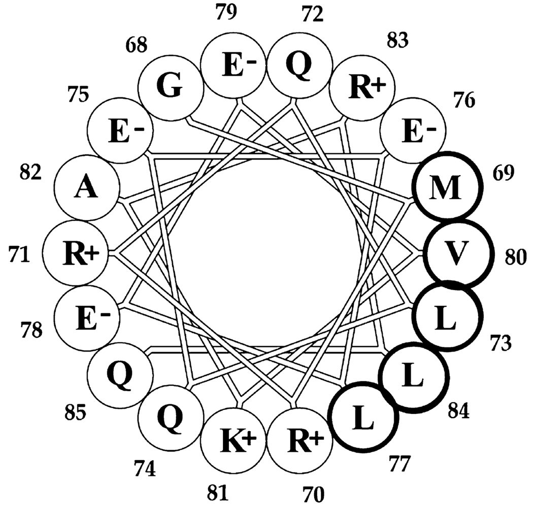
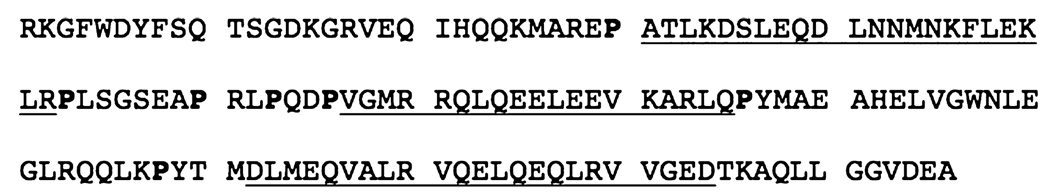
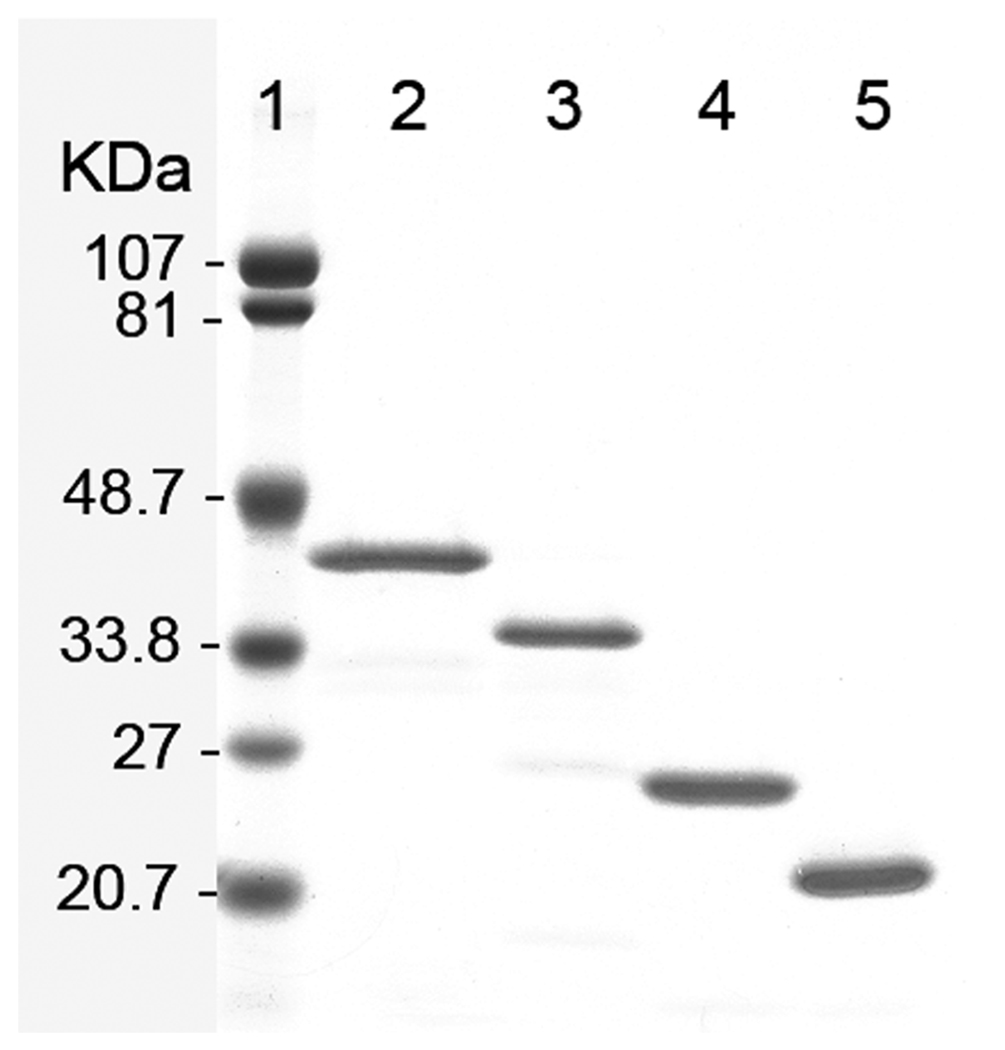
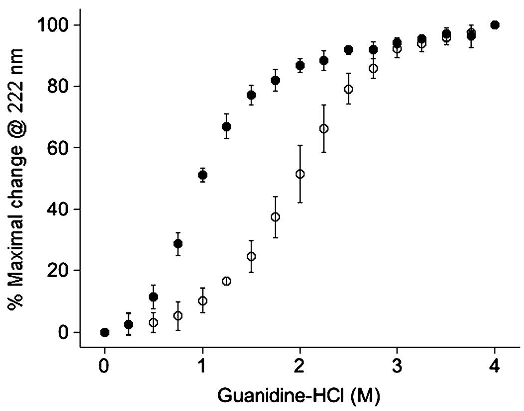
Previous studies of recombinant full-length human apolipoprotein A-V (apoA-V) provided evidence of the presence of two independently folded structural domains. Computer-assisted sequence analysis and limited proteolysis studies identified an N-terminal fragment as a candidate for one of the domains. C-Terminal truncation variants in this size range, apoA-V(1-146) and apoA-V(1-169), were expressed in Escherichia coli and isolated. Unlike full-length apoA-V or apoA-V(1-169), apoA-V(1-146) was soluble in neutral-pH buffer in the absence of lipid. Sedimentation equilibrium analysis yielded a weight-average molecular weight of 18811, indicating apoA-V(1-146) exists as a monomer in solution. Guanidine HCl denaturation experiments at pH 3.0 yielded a one-step native to unfolded transition that corresponds directly with the more stable component of the two-stage denaturation profile exhibited by full-length apoA-V. On the other hand, denaturation experiments conducted at pH 7.0 revealed a less stable structure. In a manner similar to that of known helix bundle apolipoproteins, apoA-V(1-146) induced a relatively small enhancement in 8-anilino-1-naphthalenesulfonic acid fluorescence intensity. Quenching studies with single-Trp apoA-V(1-146) variants revealed that a unique site predicted to reside on the nonpolar face of an amphipathic alpha-helix was protected from quenching by KI. Taken together, the data suggest the 146 N-terminal residues of human apoA-V adopt a helix bundle molecular architecture in the absence of lipid and, thus, likely exist as an independently folded structural domain within the context of the intact protein.
Figures


References
-
- Jakel H, Nowak M, Helleboid-Chapman A, Fruchart-Najib J, Fruchart JC. Is apolipoprotein A5 a novel regulator of triglyceride-rich lipoproteins? Ann. Med. 2006;38:2–10. - PubMed
-
- Wong K, Ryan RO. Characterization of apolipo-protein A-V structure and mode of plasma triacylglycerol regulation. Curr. Opin. Lipidol. 2007;18:319–324. - PubMed
-
- Pennacchio LA, Olivier M, Hubacek JA, Cohen JC, Cox DR, Fruchart JC, Krauss RM, Rubin EM. An apolipoprotein influencing triglycerides in humans and mice revealed by comparative sequencing. Science. 2001;294:169–173. - PubMed
-
- Alborn WE, Johnson MG, Prince MJ, Konrad RJ. Definitive N-terminal protein sequence and further characterization of the novel apolipoprotein A5 in human serum. Clin. Chem. 2006;52:514–517. - PubMed
Publication types
MeSH terms
Substances
Grants and funding
LinkOut - more resources
Full Text Sources
Miscellaneous
