. 2008 Aug 19;47(33):8527-37.
doi: 10.1021/bi800324r. Epub 2008 Jul 25.

Mechanism and dynamics of translesion DNA synthesis catalyzed by the Escherichia coli Klenow fragment

Affiliations

Mechanism and dynamics of translesion DNA synthesis catalyzed by the Escherichia coli Klenow fragment

Asim Sheriff et al. Biochemistry. .

Abstract

Translesion DNA synthesis represents the ability of a DNA polymerase to incorporate and extend beyond damaged DNA. In this report, the mechanism and dynamics by which the Escherichia coli Klenow fragment performs translesion DNA synthesis during the misreplication of an abasic site were investigated using a series of natural and non-natural nucleotides. Like most other high-fidelity DNA polymerases, the Klenow fragment follows the "A-rule" of translesion DNA synthesis by preferentially incorporating dATP opposite the noninstructional lesion. However, several 5-substituted indolyl nucleotides lacking classical hydrogen-bonding groups are incorporated approximately 100-fold more efficiently than the natural nucleotide. In general, analogues that contain large substituent groups in conjunction with significant pi-electron density display the highest catalytic efficiencies ( k cat/ K m) for incorporation. While the measured K m values depend upon the size and pi-electron density of the incoming nucleotide, k cat values are surprisingly independent of both biophysical features. As expected, the efficiency by which these non-natural nucleotides are incorporated opposite templating nucleobases is significantly reduced. This reduction reflects minimal increases in K m values coupled with large decreases in k cat values. The kinetic data obtained with the Klenow fragment are compared to that of the high-fidelity bacteriophage T4 DNA polymerase and reveal distinct differences in the dynamics by which these non-natural nucleotides are incorporated opposite an abasic site. These biophysical differences argue against a unified mechanism of translesion DNA synthesis and suggest that polymerases employ different catalytic strategies during the misreplication of damaged DNA.


Figures

Figure 1
Figure 1
(A) Structures of 2' deoxynucleoside triphosphates used or referred to in this study are dATP, dCTP, dGTP, dTTP, Ind-TP, 5-FITP, 5-AITP, 5-NITP, 5-PhITP, 5-CE-ITP, 5-CH-ITP, and 5-NapITP. For convenience, dR is used to represent the 2'-deoxyribose 5'-triphosphate portion of the nucleotides. (B) Defined DNA substrates used for kinetic analysis. “X” in the template strand denotes T or the presence of a tetrahydrofuran moiety that mimics an abasic site.
Figure 2
Figure 2
(A) Representative denaturing gel electrophoresis data for the incorporation of natural nucleotides opposite an abasic site catalyzed by the E. coli Klenow fragment. Experiments were performed mixing a preincubated solution of 60 nM Klenow fragment and 1 μM DNA with 100 μM EDTA against 100 μM nucleotide and 10 mM Mg2+. Reactions were quenched at 120 seconds by the addition of 350 mM EDTA. Quenched samples were diluted 1:1 with sequencing gel load buffer and products were analyzed for product formation by denaturing gel electrophoresis. (B) Dependency of the steady-state rate in primer elongation on dATP concentration. The following concentrations of dATP were used: 50 μM (○), 100 μM (●), 250 μM (◻), 350 μM (∎), and 500 μM (▵). The solid lines represent the fit of each set of data to a straight line. (C) The rates in primer elongation (●) were plotted versus dATP concentration and fit to the Michaelis-Menten equation to determine a Km of 146 +/- 46 μM and a Vmax of 9.6 +/- 1.0 nM/sec. The kcat value of 0.8 +/- 0.1 sec-1 was obtained by dividing Vmax by the concentration of polymerase.
Figure 3
Figure 3
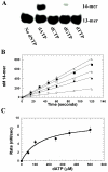
(A) Representative denaturing gel electrophoresis data for the incorporation of non-natural nucleotides opposite an abasic site catalyzed by the E. coli Klenow fragment. Experiments were performed mixing a preincubated solution of 12 nM Klenow fragment and 1 μM DNA with 100 μM EDTA against 100 μM nucleotide and 10 mM Mg2+. Reactions were quenched at 15 seconds by adding 350 mM EDTA. Quenched samples were diluted 1:1 with sequencing gel load buffer and products were analyzed for product formation by denaturing gel electrophoresis. (B) Dependency of the steady-state rate in primer elongation on 5-NITP concentration. The following concentrations of 5-NITP were used: 5 μM (○), 10 μM (●), 25 μM (◻), 50 μM (∎), 100 μM (▵), and 200 μM (▴). The solid lines represent the fit of each set of data to a linear function to determine rates. (C) The rates in primer elongation (○) were plotted versus 5-NITP concentration and fit to the Michaelis-Menten equation to determine a Km value of 74 +/- 14 μM and a Vmax of 28 +/- 2.4 nM/sec. The kcat value of 2.3 +/- 0.2 sec-1 was obtained by dividing Vmax by the concentration of polymerase.
Figure 4
Figure 4
Comparison of the active sites of the (A) KlenTaq DNA polymerase (PDB ID:3KTQ) and (B) the bacteriophage RB69 DNA polymerase (PDB ID:1IG9) (B). The active site is defined as the amino acids that lie within 6Å from the primer-template junction (shown as a space-filled model). For clarity, these amino acids are color-coded as follows: amino acids that are neutral, hydrophilic, and polar are in orange; amino acids that are positively charged and basic are in blue; amino acids that are negatively charged and acidic are in white; amino acids that are aliphatic and nonpolar are in green; and aromatic amino acids are in yellow. Swiss PDB Viewer 3.7 (www.expasy.org/spdbv) and MOE 2007.09 (www.chemcomp.com) were used to prepare these models. Insets provided in A and B show the arrangement of conserved amino acids that participate in the chemistry of phosphoryl transfer.
Figure 5
Figure 5
Comparison of the difference in the orientation of specific residues in the active sites of (A) KlenTaq DNA polymerase with respect to the docked structure of 5-NITP versus (B) the bacteriophage RB69 DNA polymerase with reference to the 5-NITP co-crystallized structure (PDB ID:2OZM).
Figure 6
Figure 6
Comparison of the docked structures of (A) 5-PhITP and (B) 5-NITP in the active site of the KlenTaq DNA polymerase. Both nucleotides are shown in ball and stick representation and in CPK color scheme for clarity. The aromatic amino acids F667 and Y671 are shown in yellow.

References

    1. Knowles JR. Enzyme-catalyzed phosphoryl transfer reactions. Annu. Rev. Biochem. 1980;49:877–919. - PubMed
    1. Kunkel TA, Bebenek K. DNA replication fidelity. Annu. Rev. Biochem. 2000;69:497–529. - PubMed
    1. Johnson KA. Conformational coupling in DNA polymerase fidelity. Annu. Rev. Biochem. 1993;62:685–713. - PubMed
    1. Goodman MF, Fygenson KD. DNA polymerase fidelity: from genetics toward a biochemical understanding. Genetics. 1998;148:1475–1482. - PMC - PubMed
    1. Beckman RA, Loeb LA. Multi-stage proofreading in DNA replication. Q. Rev. Biophys. 1993;26:225–331. - PubMed

Publication types