

Plasma levels of soluble Tie2 and vascular endothelial growth factor distinguish critical limb ischemia from intermittent claudication in patients with peripheral arterial disease
- PMID: 18652948
- PMCID: PMC2643047
- DOI: 10.1016/j.jacc.2008.02.045
Plasma levels of soluble Tie2 and vascular endothelial growth factor distinguish critical limb ischemia from intermittent claudication in patients with peripheral arterial disease
Abstract
Objectives: Our purpose was to determine whether factors that regulate angiogenesis are altered in peripheral arterial disease (PAD) and whether these factors are associated with the severity of PAD.
Background: Alterations in angiogenic growth factors occur in cardiovascular disease (CVD), but whether these factors are altered in PAD or correlate with disease severity is unknown.
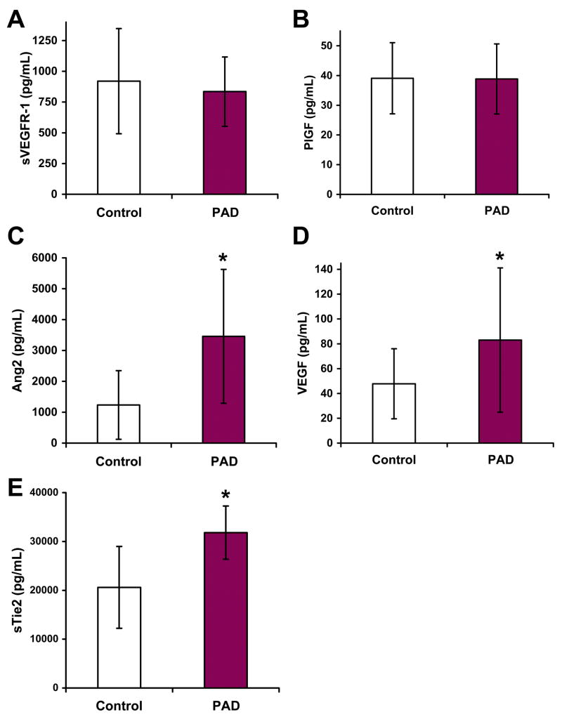
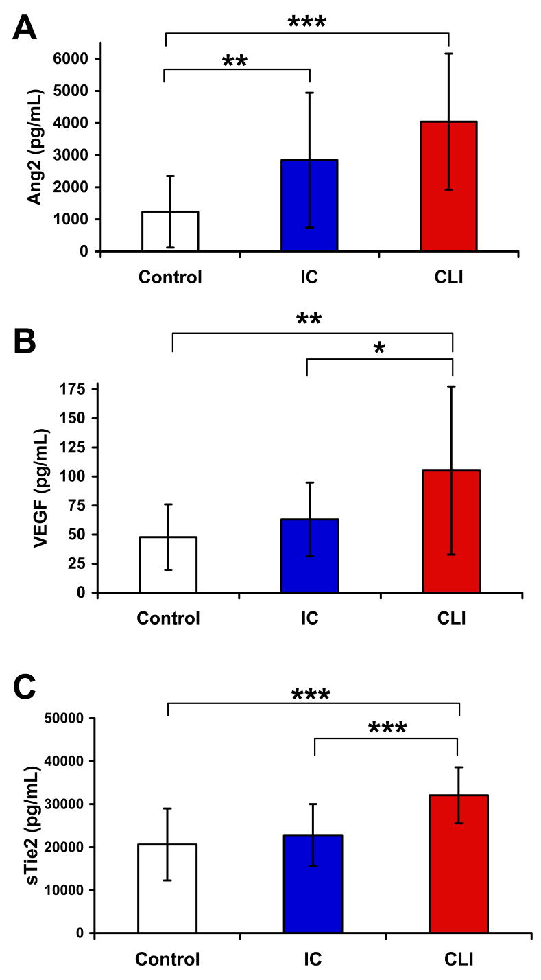
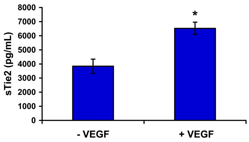
Methods: Plasma was collected from patients with PAD (n = 46) and healthy control subjects (n = 23). Peripheral arterial disease patients included those with intermittent claudication (IC) (n = 23) and critical limb ischemia (CLI) (n = 23). Plasma angiopoietin-2 (Ang2), soluble Tie2 (sTie2), vascular endothelial growth factor (VEGF), soluble VEGF receptor 1 (sVEGFR-1), and placenta growth factor (PlGF) were measured by enzyme-linked immunoadsorbent assay. In vitro, endothelial cells (ECs) were treated with recombinant VEGF to investigate effects on sTie2 production.
Results: Plasma concentrations of sTie2 (p < 0.01), Ang2 (p < 0.001), and VEGF (p < 0.01), but not PlGF or sVEGFR-1, were significantly greater in PAD patients compared with control subjects. Plasma Ang2 was significantly increased in both IC and CLI compared with control subjects (p < 0.0001), but there was no difference between IC and CLI. Plasma VEGF and sTie2 were similar in control subjects and IC but were significantly increased in CLI (p < 0.001 vs. control or IC). Increased sTie2 and VEGF were independent of CVD risk factors or the ankle-brachial index, and VEGF treatment of ECs in vitro significantly increased sTie2 shedding.
Conclusions: Levels of VEGF and sTie2 are significantly increased in CLI, and sTie2 production is induced by VEGF. These proteins may provide novel biomarkers for CLI, and sTie2 may be both a marker and a cause of CLI.
Conflict of interest statement
Conflict of Interest/Financial Disclosures: None
Figures
Comment in
-
Critical determinants of limb ischemia.J Am Coll Cardiol. 2008 Jul 29;52(5):394-6. doi: 10.1016/j.jacc.2008.05.004. J Am Coll Cardiol. 2008. PMID: 18652949 Free PMC article. No abstract available.
References
-
- Criqui MH. Peripheral arterial disease--epidemiological aspects. Vasc Med. 2001;6:3–7. - PubMed
-
- Dolan NC, Liu K, Criqui MH, et al. Peripheral artery disease, diabetes, and reduced lower extremity functioning. Diabetes Care. 2002;25:113–20. - PubMed
-
- Ouriel K. Peripheral arterial disease. Lancet. 2001;358:1257–64. - PubMed
-
- Weitz JI, Byrne J, Clagett GP, et al. Diagnosis and treatment of chronic arterial insufficiency of the lower extremities: a critical review. Circulation. 1996;94:3026–49. - PubMed
Publication types
MeSH terms
Substances
Grants and funding
LinkOut - more resources
Full Text Sources
Other Literature Sources
Research Materials
Miscellaneous
