. 2008 Sep;87(3):208-13.
doi: 10.1016/j.exer.2008.05.017. Epub 2008 Jun 3.

Inflammatory demyelination induces axonal injury and retinal ganglion cell apoptosis in experimental optic neuritis

Affiliations

Inflammatory demyelination induces axonal injury and retinal ganglion cell apoptosis in experimental optic neuritis

Kenneth S Shindler et al. Exp Eye Res. 2008 Sep.

Abstract

Optic neuritis is an inflammatory disease of the optic nerve that often occurs in patients with multiple sclerosis and leads to permanent visual loss mediated by retinal ganglion cell (RGC) damage. Optic neuritis occurs with high frequency in relapsing-remitting experimental autoimmune encephalomyelitis (EAE), an animal model of multiple sclerosis, with significant loss of RGCs. In the current study, mechanisms of RGC loss in this model were examined to determine whether inflammation-induced axonal injury mediates apoptotic death of RGCs. RGCs were retrogradely labeled by injection of fluorogold into superior colliculi of 6-7 week old female SJL/J mice. EAE was induced one week later by immunization with proteolipid protein peptide. Optic neuritis was detected by inflammatory cell infiltration on histological examination as early as 9 days after immunization, with peak incidence by day 12. Demyelination occurred 1-2 days after inflammation began. Loss of RGC axons was detected following demyelination, with significant axonal loss occurring by day 13 post-immunization. Axonal loss occurred prior to loss of RGC bodies at day 14. Apoptotic cells were also observed at day 14 in the ganglion cell layer of eyes with optic neuritis, but not in control eyes. Together these results suggest that inflammatory cell infiltration mediates demyelination and leads to direct axonal injury in this model of experimental optic neuritis. RGCs die by an apoptotic mechanism triggered by axonal injury. Potential neuroprotective therapies to prevent permanent RGC loss from optic neuritis will likely need to be initiated prior to axonal injury to preserve neuronal function.


Figures

Fig. 1
Fig. 1
EAE optic nerves develop demyelination. (A) H & E stained section of optic nerve from a day 14 control mouse showing normal histology. (B) Inflammatory cell infiltrates observed in the optic nerve (between arrowheads) and optic nerve sheath (arrow) from a day 14 EAE mouse with optic neuritis. (C) Higher magnification view of inflammatory cells in the nerve shown in B. (D) Luxol fast blue staining in the control optic nerve shown in A demonstrates the normal myelinated nerve. (E) The EAE optic nerve shown in B contains an area of normal myelin (arrowhead) along with mild diffuse reduction in staining and focal areas of complete demyelination (arrows). (F) Higher magnification view of an area of demyelination (arrow). All scale bars = 50 μm.
Fig. 2
Fig. 2
Optic nerve demyelination begins after onset of inflammation and progresses over several days. (A) The earliest time optic nerve inflammation was detected in EAE eyes was 9 days post-immunization. 33.3% incidence of inflammation at day 9 increased through day 12 and remained steady at 64.3% (9 of 14 eyes) through day 14. In contrast, no demyelination was detected until day 11, and the incidence of demyelination increased to 57.2% (8 of 14 eyes) by day 14. (B) The severity of demyelination, scored on a 0–3 point scale, also increased over time. The average score ± SEM per eye with demyelination is shown. One representative experiment of three is shown.
Fig. 3
Fig. 3
Injury and loss of axons in day 14 EAE optic nerves. (A) Normal density of RGC axons observed with silver staining in a control optic nerve. (B) An optic nerve from a day 14 EAE mouse demonstrates reduced axonal staining and disorganization from axonal injury. (C) Higher magnification view of injured nerve in B with truncated axon (arrowhead) and terminal axonal ovoid swelling (arrow). (D) EAE eyes that developed optic neuritis (10 of 15 eyes) had a significant reduction in the area of axonal staining (9558 ± 1357 μm2) compared with control eyes from sham-immunized mice (19104 ± 471; *p ≤ 0.01). EAE eyes that did not develop optic neuritis had no significant decrease in axonal area (15998 ± 3489) compared to controls. Data represent the mean ± SEM. One representative experiment of three is shown. Scale bars (A–C) = 50 μm.
Fig. 4
Fig. 4
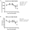
Axonal loss precedes RGC loss. (A) The average axonal area (mean ± SEM) measured from days 7 –14 in control and EAE optic nerves with or without optic neuritis is shown. Control nerves from age-matched normal mice (no CFA) and from mice sham-immunized with CFA were used. Significant reduction in axonal staining in optic neuritis vs. control (+/− CFA) eyes is first detected at day 13 (*p ≤ 0.05) and persists at day 14 (**p ≤ 0.01). 5 – 10 nerves were examined per group. (B) The number of surviving RGCs (mean ± SEM) in the retina of EAE eyes with optic neuritis declines slowly, with no significant difference compared to control eye RGC numbers detected until day 14 (*p ≤ 0.05). Retinas of 5 – 6 eyes were counted per group. Data represent results from one of three experiments.
Fig. 5
Fig. 5
Apoptosis of RGCs in optic neuritis eyes. (A) H & E stained section through a control eye shows the normal single layer of RGC nuclei in the ganglion cell layer. (B) No TUNEL positive cells were detected in the control eye shown in A. (C,D) Arrows indicate two RGC nuclei with condensed chromatin that were undergoing apoptosis as indicated by positive TUNEL labeling. Scale bars = 50 μm.

References

    1. Arnold AC. Evolving management of optic neuritis and multiple sclerosis. Am J Ophthalmol. 2005;139:1101–1108. - PubMed
    1. Beck RW, Cleary PA, Anderson MM, Jr, Keltner JL, Shults WT, Kaufman DI, Buckley EG, Corbett JJ, Kupersmith MJ, Miller NR, et al. A randomized, controlled trial of corticosteroids in the treatment of acute optic neuritis. New Eng J Med. 1992;326:581–588. - PubMed
    1. Berkelaar M, Clarke DB, Wang YC, Bray GM, Aguayo AJ. Axotomy results in delayed death and apoptosis of retinal ganglion cells in adult rats. J Neurosci. 1994;14:4368–4374. - PMC - PubMed
    1. Bettelli E, Pagany M, Weiner HL, Linington C, Sobel RA, Kuchroo VK. Myelin oligodendrocyte glycoprotein-specific T cell receptor transgenic mice develop spontaneous autoimmune optic neuritis. J Exp Med. 2003;197:1073–81. - PMC - PubMed
    1. Costello F, Coupland S, Hodge W, Lorello GR, Koroluk J, Pan YI, Freedman MS, Zackon DH, Kardon RH. Quantifying axonal loss after optic neuritis with optical coherence tomography. Ann Neurol. 2006;59:963–969. - PubMed

Publication types