. 2008 Oct;82(19):9359-68.
doi: 10.1128/JVI.00242-08. Epub 2008 Jul 23.

Transcriptional control of spliced and unspliced human T-cell leukemia virus type 1 bZIP factor (HBZ) gene

Affiliations

Transcriptional control of spliced and unspliced human T-cell leukemia virus type 1 bZIP factor (HBZ) gene

Mika Yoshida et al. J Virol. 2008 Oct.

Abstract

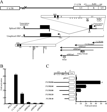
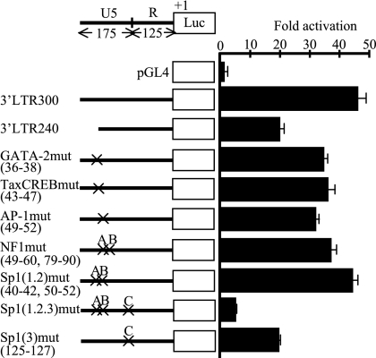
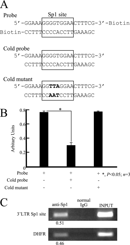
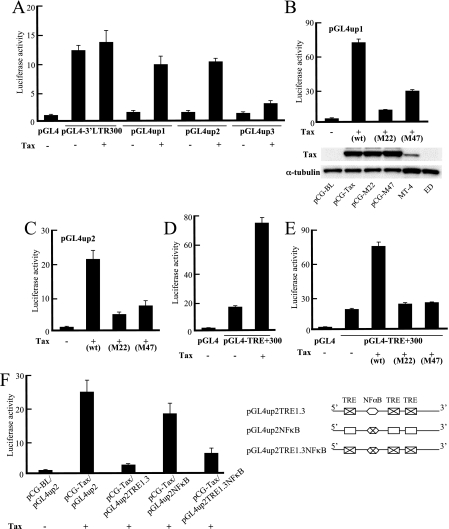
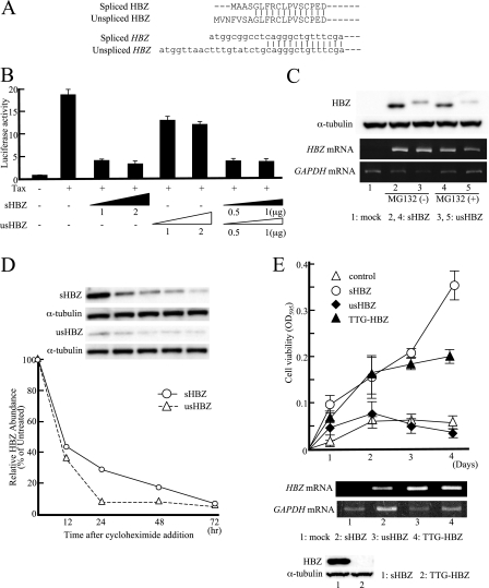
The human T-cell leukemia virus type 1 (HTLV-1) basic leucine zipper factor (HBZ) gene is encoded by the minus strand of the HTLV-1 provirus and transcribed from the 3' long terminal repeat (LTR). HBZ gene expression not only inhibits the Tax-mediated activation of viral gene transcription through the 5' LTR but also promotes the proliferation of infected cells. However, the HBZ promoter region and the transcriptional regulation of the gene have not been studied. In this study, we characterize the promoters of the spliced version of the HBZ gene (sHBZ) and the unspliced version of the HBZ gene (usHBZ) by luciferase assay. Both promoters were TATA-less and contained initiators and downstream promoter elements. Detailed studies of the promoter for the sHBZ gene showed that Sp1 sites were critical for its activity. The activities of the sHBZ and usHBZ gene promoters were upregulated by Tax through Tax-responsible elements in the 3' LTR. We compared the functions of the proteins derived from the sHBZ and usHBZ transcripts. sHBZ showed a stronger suppression of Tax-mediated transcriptional activation through the 5' LTR than did usHBZ; the level of suppression correlated with the level of protein produced. The expression of sHBZ had a growth-promoting function in a T-cell line, while usHBZ expression did not. This study demonstrates that Sp1 is critical for sHBZ transcription, which accounts for the constitutive expression of the sHBZ gene. Functional differences between sHBZ and usHBZ suggest that the sHBZ gene plays a significant role in the proliferation of infected cells.


Figures

FIG. 1.
FIG. 1.
Characterization of the HBZ promoter. (A) Identification of transcriptional start sites of usHBZ by 5′-RACE. Schematic diagrams show the HTLV-1 provirus, the sHBZ and usHBZ transcripts, and the expected promoter regions that we subcloned for analysis. The asterisks show identified transcriptional initiation sites of sHBZ and usHBZ transcripts. The expected promoter region of sHBZ (3′LTR300) consists of a 300-bp segment containing 175 bp from the U5 region and 125 bp from the R region, while the promoter for usHBZ is believed to lie somewhere upstream of the usHBZ coding sequence (uHBZup1, uHBZup2, and uHBZup3). 3′LTRwhole contains the whole LTR sequence. (B) Comparison of sHBZ and usHBZ promoter activities. Reporter constructs containing the promoter segments shown in A were transiently transfected into Jurkat cells (2 × 10⁵ cells) and assayed for luciferase activity. The luciferase activity relative to that in the cell lysate transfected by empty vector (pGL4) is shown. Values represent means ± SD (n = 3). (C) Luciferase activities of deletion mutants of the sHBZ promoter. A reporter plasmid containing a DNA segment upstream of the transcription initiation sites (full length [300 bp] or a deletion construct [1 μg each]) was transfected into Jurkat cells (2 × 10⁵ cells). After 2 days, luciferase activity was measured. The luciferase activity relative to that of cells transfected with empty vector (pGL4.22) is shown. Values represent means ± SD (n = 3).
FIG. 2.
FIG. 2.
Mutation analysis of the sHBZ promoter. A reporter construct (1 μg) containing the full-length sHBZ promoter (pGL4-3′LTR300), a 60-bp deletion mutant (pGL4-3′LTR240), or one of seven other mutated constructs was transfected into Jurkat cells (2 × 10⁵ cells), and luciferase activity (relative to that of an empty vector control) was measured after 2 days. Each × represents the position of a site-directed mutation in a transcriptional factor recognition site. Each mutation resulted in a loss of consensus sequence. Values represent means ± SD (n = 3).
FIG. 3.
FIG. 3.
Binding of Sp1 to the 3′ LTR. (A) Oligonucleotides used to study the binding activities of Sp1. Double-stranded biotinylated or unbiotinylated oligonucleotides corresponding to the Sp1 site in the sHBZ promoter were synthesized. The biotinylated wild-type probe (Probe), the competing nonbiotinylated oligonucleotide (Cold probe), and an oligonucleotide modified to contain a mutation in the Sp1 recognition sequence (Cold mutant) (mutated bases are in boldface type) are shown. (B) NoShift assay to detect Sp1 binding activity. The activity of Sp1 binding to the recognition site was abolished by excess cold (nonbiotinylated) oligonucleotide but not by excess mutant oligonucleotide (*, P = 0.026). (C) ChIP assay with anti-Sp1 antibody. Formaldehyde-cross-linked chromatin was isolated from ATL-43T cells. Chromatin was immunoprecipitated with anti-Sp1 or normal rabbit immunoglobulin G (IgG) (as a negative control). Immunoprecipitated DNA was purified and analyzed by PCR using primers specific for the 3′ LTR Sp1 site or the DHFR gene (positive control for anti-Sp1 ChIP). Relative values (each input band density equals 1) are shown below anti-Sp1 bands.
FIG. 4.
FIG. 4.
Effects of Tax or HBZ on the sHBZ promoter. (A) Effect of Tax on the sHBZ promoter. Jurkat cells (2 × 10⁵ cells) were cotransfected with 1 μg each of pCG-Tax and pGL4-3′LTR300 and assayed for luciferase activity. The total amount of DNA to be introduced was equalized with empty vector (pCG-BL or pGL4.22). Activation relative to that of cells transfected with pCG-Tax and pGL4.22 is shown. (B) Effect of HBZ on the sHBZ promoter. Jurkat cells (2 × 10⁵ cells) were cotransfected with 1 μg each of pME18SneoHBZ and pGL4-3′LTR300 and assayed for luciferase activity. The total amount of DNA to be introduced was equalized with empty vector (pME18Sneo or pGL4.22). Activation relative to that of cells transfected with pME18sneoHBZ and pGL4.22 is shown. Values represent means ± SD (n = 3).
FIG. 5.
FIG. 5.
Tax-mediated transactivation of the usHBZ and sHBZ gene promoters. Jurkat cells were cotransfected with 1 μg of wild-type (wt) or mutated Tax expression vector and 1 μg of sHBZ or usHBZ promoter luciferase reporter constructs. Luciferase activities were measured. Activation relative to that of cells transfected with pCG-BL and each fundamental vector (pGL4.22 [A, D, and E], pGL4up1 [B], and pGL4up2 [C and F]) is shown. Values represent means ± SD (n = 3). (A) Tax-mediated transactivation of the usHBZ promoter deletion mutants (up1 and up2). (B and C) Effects of Tax mutants (M22 and M47) on the promoter of usHBZ (B, up1; C, up2). (B, bottom) Western blot analysis of Tax in 293FT cells transfected pGL4up1 and each tax (wild type, M22, or M47) expression vector. MT-4 was used as the positive control. (D) Tax-mediated transactivation of the construct that contains both the sHBZ promoter and the TRE sequence. (E) Effects of Tax mutants (M22 and M47) on the TREs and sHBZ promoter. (F) Effects of mutations in the usHBZ promoter (up2). Constructs similar to up2, but with mutated TREs and/or a mutated NF-κB site, were made and tested as shown.
FIG. 6.
FIG. 6.
Functional differences between sHBZ and usHBZ. (A) Nucleotide and peptide sequences at the N terminus of the two HBZ isoforms are shown. (B) Different functions of the sHBZ and usHBZ proteins in Tax-mediated viral gene transactivation. Jurkat cells were cotransfected with 1 μg of pCG-Tax and either pME18Sneo-splicedHBZ (lane 3, 1 μg; lane 4, 2 μg), pME18Sneo-unsplicedHBZ (lane 5, 1 μg; lane 6, 2 μg), or both (lane 7, 0.5 μg each; lane 8, 1 μg each) and assayed for luciferase activity. The total amount of DNA to be introduced was equalized with empty vector (pCG-BL and pME18Sneo). After 2 days, cells were lysed, and relative luciferase activities were determined. Activation relative to that of cells transfected with pCG-BL, pME18Sneo, and pGL4.22 is shown. Values represent means ± SD (n = 3). (C) Expression levels of protein and mRNAs of the two HBZ isoforms. Cell extracts were prepared from 293FT cells transfected with pcDNA3.1(−), pcDNA3.1(−)sHBZ-MycHis, or pcDNA3.1(−)usHBZ-MycHis. HBZ protein isoforms were detected by Western blotting using anti-His antibodies. At the same time, the transcription of the two HBZ isoforms was studied by semiquantitative RT-PCR. To evaluate whether differences in proteasome-dependent degradation contributed to the differences in HBZ expression levels, some samples were treated with MG132 before extraction of protein or mRNA. Lane 1, mock; lanes 2 and 4, sHBZ transfectants; lanes 3 and 5, usHBZ transfectants. (D) Stability of sHBZ and usHBZ proteins by cycloheximide pulse-chase assay. Forty-eight hours after transfection, 293FT cells were treated with cycloheximide (100 μg/ml) for the indicated times, followed by an immunoblotting assay with an anti-His antibody or an anti-tubulin antibody. Densitometric quantification of sHBZ (full line) and usHBZ (dashed line) is shown, with the quantification of each HBZ isoform at 0 h after cycloheximide treatment defined as 100%, upon which the change in expression levels was calculated. (E) Functional differences of sHBZ, usHBZ, and TTG-HBZ. The effects of the two HBZ isoform genes on Kit225 cell growth were measured as described in Materials and Methods. Values represent means ± standard deviations (error bars) (n = 3). Three independent experiments, each with triplicate transfections, were performed, and typical results are shown. (Bottom) Transcription of HBZ was analyzed by semiquantitative RT-PCR. Lane 1, mock; lanes 2, sHBZ transfectant; lane 3, usHBZ transfectant; lane 4, TTG-HBZ transfectant. (Bottom) The TTG-HBZ protein was analyzed by the Western blot method. Lane 1, sHBZ transfectant; lane 2, TTG-HBZ transfectant.

References

    1. Arnold, J., B. Yamamoto, M. Li, A. J. Phipps, I. Younis, M. D. Lairmore, and P. L. Green. 2006. Enhancement of infectivity and persistence in vivo by HBZ, a natural antisense coded protein of HTLV-1. Blood 1073976-3982. - PMC - PubMed
    1. Basbous, J., C. Arpin, G. Gaudray, M. Piechaczyk, C. Devaux, and J. M. Mesnard. 2003. The HBZ factor of human T-cell leukemia virus type I dimerizes with transcription factors JunB and c-Jun and modulates their transcriptional activity. J. Biol. Chem. 27843620-43627. - PubMed
    1. Boam, D. S., I. Davidson, and P. Chambon. 1995. A TATA-less promoter containing binding sites for ubiquitous transcription factors mediates cell type-specific regulation of the gene for transcription enhancer factor-1 (TEF-1). J. Biol. Chem. 27019487-19494. - PubMed
    1. Cavanagh, M. H., S. Landry, B. Audet, C. Arpin-Andre, P. Hivin, M. E. Pare, J. Thete, E. Wattel, S. J. Marriott, J. M. Mesnard, and B. Barbeau. 2006. HTLV-I antisense transcripts initiating in the 3′ LTR are alternatively spliced and polyadenylated. Retrovirology 315. - PMC - PubMed
    1. Datta, S., N. H. Kothari, and H. Fan. 2000. In vivo genomic footprinting of the human T-cell leukemia virus type 1 (HTLV-1) long terminal repeat enhancer sequences in HTLV-1-infected human T-cell lines with different levels of Tax I activity. J. Virol. 748277-8285. - PMC - PubMed

Publication types

MeSH terms

Substances

LinkOut - more resources