. 2008 Oct;19(10):4130-40.
doi: 10.1091/mbc.e07-11-1146. Epub 2008 Jul 23.

A mitotic GlcNAcylation/phosphorylation signaling complex alters the posttranslational state of the cytoskeletal protein vimentin

Affiliations

A mitotic GlcNAcylation/phosphorylation signaling complex alters the posttranslational state of the cytoskeletal protein vimentin

Chad Slawson et al. Mol Biol Cell. 2008 Oct.

Abstract

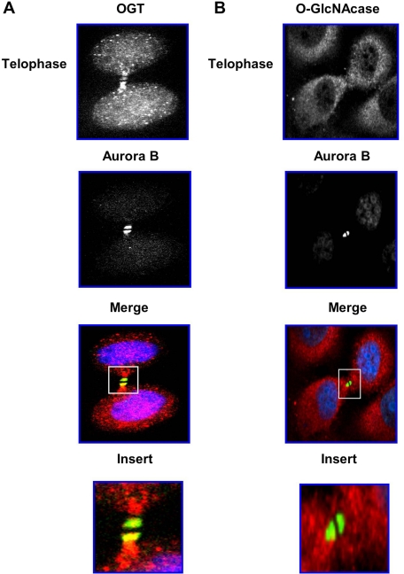
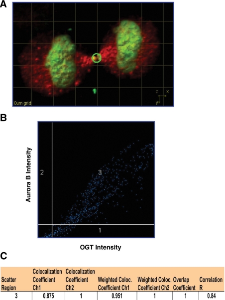
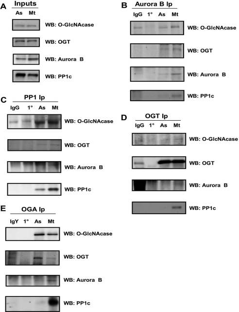
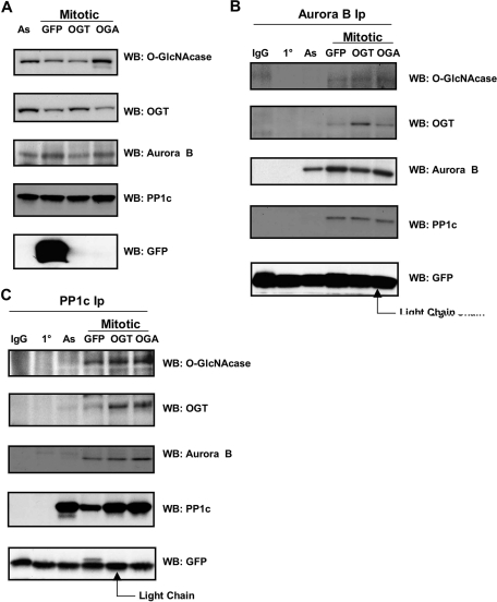
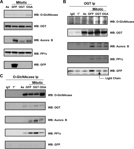
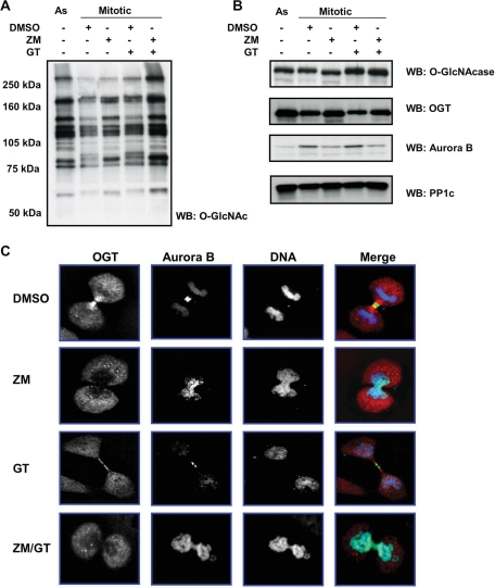
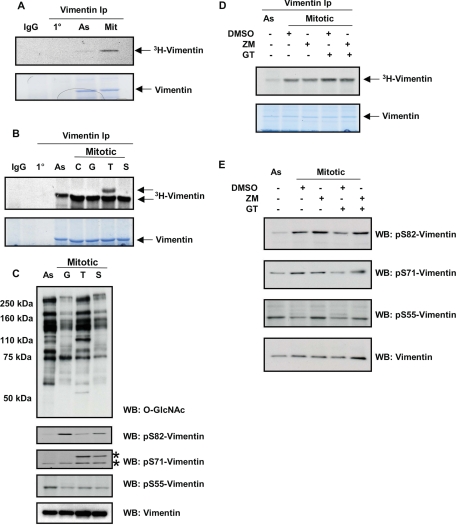
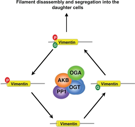
O-linked beta-N-acetylglucosamine (O-GlcNAc) is a highly dynamic intracellular protein modification responsive to stress, hormones, nutrients, and cell cycle stage. Alterations in O-GlcNAc addition or removal (cycling) impair cell cycle progression and cytokinesis, but the mechanisms are not well understood. Here, we demonstrate that the enzymes responsible for O-GlcNAc cycling, O-GlcNAc transferase (OGT) and O-GlcNAcase (OGA) are in a transient complex at M phase with the mitotic kinase Aurora B and protein phosphatase 1. OGT colocalized to the midbody during telophase with Aurora B. Furthermore, these proteins coprecipitated with each other in a late mitotic extract. The complex was stable under Aurora inhibition; however, the total cellular levels of O-GlcNAc were increased and the localization of OGT was decreased at the midbody after Aurora inhibition. Vimentin, an intermediate filament protein, is an M phase substrate for both Aurora B and OGT. Overexpression of OGT or OGA led to defects in mitotic phosphorylation on multiple sites, whereas OGT overexpression increased mitotic GlcNAcylation of vimentin. OGA inhibition caused a decrease in vimentin late mitotic phosphorylation but increased GlcNAcylation. Together, these data demonstrate that the O-GlcNAc cycling enzymes associate with kinases and phosphatases at M phase to regulate the posttranslational status of vimentin.


Figures

Figure 1.
Figure 1.
O-GlcNAc transferase localizes with Aurora B at the midbody. (A and B) HeLa cells were synchronized into G1/S by double thymidine block and released. Cells were fixed at 12 h after release. Cells were triply labeled at telophase. OGT is red, Aurora B green, and DNA blue. (A) Endogenous OGT and Aurora B strongly costain at the midbody. (B) Endogenous OGA stains diffusely throughout the cell, and only a small amount was found at the midbody compared with Aurora B. All costaining experiments were performed a minimum of three times.
Figure 2.
Figure 2.
Quantification of three-dimensional colocalization of Aurora B and O-GlcNAc transferase. (A) Three-dimensional image of a cell at telophase captured on 3i-spinning disk confocal microscope. Aurora B staining is green and OGT is red. The green circle is the area used in the quantification analysis. (B) Scatter diagram of the signal from both Aurora B and OGT. Area 3 of the diagram is the area of colocalization with a linear distribution of spots indicating greater colocalization. (C) The statistical analysis of fluorescent signals showed a high degree of colocalization. The overlap coefficient was 1, whereas the more stringent Pearson's analysis (Correlation R) has an overlap of 0.84 (a perfect correlation is 1, whereas no correlation is −1).
Figure 3.
Figure 3.
Endogenous Aurora B, PP1, OGT, and OGA all precipitate together in mitotic cells. (A–E) HeLa cells were synchronized by double thymidine block into G1/S and serum released. At 10 h after release, mitotic cells were collected by shake-off and replated. The cells were harvested 1 h after replating to produce a late M phase extract. Cells were lysed and used in coimmunoprecipitation studies. (A) Protein expression of OGA, OGT, Aurora B, and PP1 (30 μg of lysate per lane) as determined by Western blotting (As, asynchronous; Mt, mitotic). (B–E) Endogenous Aurora B, OGA, OGT, and PP1 precipitate each other from synchronized cells (3 mg/ml lysate was used in each precipitation, PP1 IPs were performed with the sc-443 antibody, whereas Aurora B Western blots were performed with the Rockland antibody). Nonspecific IgG and primary antibody (1°) were used as antibody controls. All immunoprecipitations were repeated a minimum of three times.
Figure 4.
Figure 4.
Overexpression of OGT and OGA does not alter the ability of Aurora B and PP1 to precipitate the mitotic complex. (A) Input lanes from samples used in immunoprecipitation studies. Cells were synchronized into M phase by nocodazole treatment and harvested 1 h after nocodazole washout. Protein expression of OGA, OGT, Aurora B, and PP1c (30 μg of lysate per lane) was determined by Western blotting (As, asynchronous). (B and C) Aurora B or PP1c were immunoprecipitated from 2 mg/ ml lysate (PP1 IPs were performed with the sc-7482 antibody). Endogenous OGT and OGA copurify with Aurora B and PP1c (GFP lane) in M phase extracts but not in asynchronous extracts. Overexpression of OGT or OGA did not alter their ability to copurify with Aurora B or PP1c. GFP was used as an infection control, whereas nonspecific IgG and primary antibody (1°) alone were used as antibody controls. All immunoprecipitations were repeated a minimum of three times.
Figure 5.
Figure 5.
Overexpression of OGT and OGA does not alter the ability of OGT and OGA to precipitate the mitotic complex. (A) Input lanes from samples used in OGT/OGA immunoprecipitation studies. Cells were synchronized into M phase by nocodazole treatment and harvested 1 h after nocodazole washout. Protein expression of OGA, OGT, Aurora B, and PP1c (30 μg of lysate per lane) was determined by Western blotting (As, asynchronous). (B and C) Endogenous OGT and OGA precipitate both Aurora B and PP1c in mitotic extracts (GFP lane; 2 mg/ml). No change in purification was seen in OGT/OGA overexpressing cells (OGA/OGT lanes). GFP was used as an infection control, whereas nonspecific IgG or IgY and primary antibody (1°) alone were used as antibody controls. All immunoprecipitations were repeated a minimum of three times.
Figure 6.
Figure 6.
Aurora B inhibition disrupts GlcNAcylation and OGT localization at M phase. Cells were synchronized by nocodazole and released for 1 h. Inhibitors were added at the time of release. (A) Western blot analysis of GlcNAcylated proteins after treatment with DMSO (control), ZM (20 μM), and GT (10 μM). O-GlcNAc levels were elevated in response to ZM, GT, and ZM/GT treatment compared with DMSO controls. (B) Western blot analysis of mitotic proteins (30 μg per lane). ZM treated cells showed a decrease in Aurora B levels and an increase in OGT levels. GT treatment decreased OGT levels and raised OGA protein levels. PP1c was unchanged by treatment. (C) Confocal staining of OGT and Aurora B after double thymidine block and release. Cells were fixed after 12 h. Inhibitors were added after 8 h of release. OGT stained red, Aurora B was green, and DNA was blue. ZM-treated cells caused a marked reduction in OGT staining at the midbody. GT appeared to have little to no effect on localization. Each experiment was done a minimum of three times.
Figure 7.
Figure 7.
Vimentin is a mitotic substrate of an O-GlcNAc/phosphate complex. (A) Galactosyltransferase labeling of precipitated vimentin from asynchronous (As) and nocodazole released mitotic extracts (Mit) with [3H]galactose. Vimentin was significantly more GlcNAcylated at M phase (top), whereas the amount precipitated was the same (bottom). (B) Vimentin was precipitated and radiolabeled as before from extracts overexpressing OGA and OGT (As, asynchronous; C, uninfected mitotic control; G, GFP overexpression control; T, O-GlcNAc transferase overexpression; S, OGA overexpression). OGT overexpression caused a new GlcNAcylated band to appear. (C) Overexpression of OGT or OGA altered the GlcNAcylation of mitotic proteins and disrupted specific mitotic phosphorylations of vimentin in cells nocodazole released. Asterisk denotes position of pS71-Vimentin. (D) GT treatment increased mitotic GlcNAcylation of vimentin (top), whereas ZM had no effect. Vimentin precipitated equally under all conditions (bottom). (E) Both ZM and GT treatment altered the mitotic phosphorylation of vimentin as judged by Western blot analysis on cell extracts (30 μg loaded per lane). Each experiment was repeated a minimum of three times.
Figure 8.
Figure 8.
An O-GlcNAc/phosphate complex forms in M phase to regulate the posttranslational state of proteins. A model representing the protein complex of Aurora kinase B (AKB), Protein Phosphatase 1 (PP1), O-GlcNAc transferase (OGT), and O-GlcNAcase (OGA) is proposed; this complex potentially regulates the posttranslational state of target proteins such as vimentin at M phase (P, phosphorylation; G, GlcNAcylation).

Comment in

  • Mol Biol Cell. 19:4019.

References

    1. Adams R. R., Maiato H., Earnshaw W. C., Carmena M. Essential roles of Drosophila inner centromere protein (INCENP) and aurora B in histone H3 phosphorylation, metaphase chromosome alignment, kinetochore disjunction, and chromosome segregation. J. Cell Biol. 2001;153:865–880. - PMC - PubMed
    1. Brickley K., Smith M. J., Beck M., Stephenson F. A. GRIF-1 and OIP106, members of a novel gene family of coiled-coil domain proteins: association in vivo and in vitro with kinesin. J. Biol. Chem. 2005;280:14723–14732. - PubMed
    1. Carmena M., Earnshaw W. C. The cellular geography of aurora kinases. Nat. Rev. Mol. Cell Biol. 2003;4:842–854. - PubMed
    1. Cetinbas N., Macauley M. S., Stubbs K. A., Drapala R., Vocadlo D. J. Identification of Asp174 and Asp175 as the key catalytic residues of human O-GlcNAcase by functional analysis of site-directed mutants. Biochemistry. 2006;45:3835–3844. - PubMed
    1. Cheung W. D., Hart G. W. AMP-activated protein kinase and p38 MAPK activate O-GlcNAcylation of neuronal proteins during glucose deprivation. J. Biol. Chem. 2008;283:13009–13020. - PMC - PubMed

Publication types