

Essential head tremor is associated with cerebellar vermis atrophy: a volumetric and voxel-based morphometry MR imaging study
- PMID: 18653686
- PMCID: PMC8118768
- DOI: 10.3174/ajnr.A1190
Essential head tremor is associated with cerebellar vermis atrophy: a volumetric and voxel-based morphometry MR imaging study
Abstract
Background and purpose: Our aim was to investigate the presence of brain gray matter (GM) abnormalities in patients with different forms of essential tremor (ET).
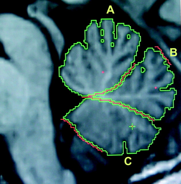
Materials and methods: We used optimized voxel-based morphometry (VBM) and manually traced single region-of-interest analysis in 50 patients with familial ET and in 32 healthy subjects. Thirty patients with ET had tremor of the arms (a-ET), whereas the remaining 20 patients had both arm and head tremor (h-ET).
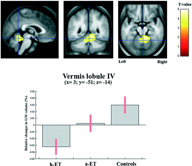
Results: VBM showed marked atrophy of the cerebellar vermis in the patients with h-ET with respect to healthy subjects (P(corrected) < .001). Patients with a-ET showed a trend toward a vermal GM volume loss that did not reach a significant difference with respect to healthy controls (P(uncorrected) < .01). The region-of-interest analysis showed a reduction of the cerebellar volume (CV) in the h-ET group (98.2 +/- 13.6 mm(3)) compared with healthy controls (110.5 +/- 15.5 mm(3), P < .012) as well as in the entire vermal area (790.3 +/- 94.5 mm(2), 898.6 +/- 170.6 mm(2), P < .04 in h-ET and control groups, respectively).
Conclusions: Atrophy of the cerebellar vermis detected in patients with h-ET strongly supports the evidence for the involvement of the cerebellum in the pathophysiology of ET. The lack of a significant CV loss observed in patients with a-ET suggests that a-ET and h-ET might represent distinct subtypes of the same disease.
Figures
References
-
- Hallett M, Dubinsky RM. Glucose metabolism in the brain of patients with essential tremor. J Neurol Sci 1993;114:45–48 - PubMed
-
- Boecker H, Brooks DJ. Functional imaging of tremor. Mov Disord 1998;13:64–72 - PubMed
-
- Louis ED, Shungu D, Chan S, et al. Metabolic abnormality in essential tremor: a magnetic resonance spectroscopic imaging study. Neurosci Lett 2002;333:17–20 - PubMed
-
- Daniels C, Peller M, Wolff S, et al. Voxel-based morphometry shows no decreases in cerebellar gray matter volume in essential tremor. Neurology 2006;67:1452–56 - PubMed
-
- Louis ED, Vonsattel JP, Honig LS, et al. Essential tremor associated with pathologic changes in the cerebellum. Arch Neurol 2006;63:1189–93 - PubMed
MeSH terms
LinkOut - more resources
Full Text Sources
Medical