. 2008 Nov;149(11):5549-56.
doi: 10.1210/en.2008-0618. Epub 2008 Jul 24.

The luteinizing hormone receptor-activated extracellularly regulated kinase-1/2 cascade stimulates epiregulin release from granulosa cells

Affiliations

The luteinizing hormone receptor-activated extracellularly regulated kinase-1/2 cascade stimulates epiregulin release from granulosa cells

Nebojsa Andric et al. Endocrinology. 2008 Nov.

Abstract

We examine the pathways involved in the luteinizing hormone receptor (LHR)-dependent activation of the epidermal growth factor (EGF) network using cocultures of LHR-positive granulosa cells and LHR-negative test cells expressing an EGF receptor (EGFR)-green fluorescent protein fusion protein. Activation of the LHR in granulosa cells results in the release of EGF-like growth factors that are detected by measuring the phosphorylation of the EGFR-green fluorescent protein expressed only in the LHR-negative test cells. Using neutralizing antibodies and real-time PCR, we identified epiregulin as the main EGF-like growth factor produced upon activation of the LHR expressed in immature rat granulosa cells, and we show that exclusive inhibition or activation of the ERK1/2 cascade in granulosa cells prevents or enhances epiregulin release, respectively, with little or no effect on epiregulin expression. These results show that the LHR-stimulated ERK1/2 pathway stimulates epiregulin release.


Figures
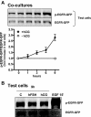
Figure 1
Figure 1
hCG-induces trans-phosphorylation of the EGFR in test cells when cocultured with granulosa cells expressing the hLHR. A, Cocultures of test cells expressing the EGFR-GFP and primary granulosa cells expressing the hLHR were incubated with buffer or hCG (100 ng/ml) for the indicated times before measuring phosphorylated and total EGFR-GFP in the lysates. The blots shown are results of a representative experiment whereas the graphs show the average ± sem of four independent experiments. B, Cultures of test cells expressing the EGFR-GFP were incubated with buffer only (C), with 100 ng/ml hCG or hFSH for 9 h, or with 100 ng/ml EGF for 15 min as indicated. The blot shown is representative of four experiments with similar results. We have previously noted a reduction in the level of total EGFR after stimulation with EGF (28). This is likely due to EGF-induced internalization and degradation of the EGFR.
Figure 2
Figure 2
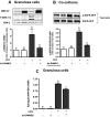
The selective inhibition of ERK1/2 phosphorylation in granulosa cells blocks the hCG-induced trans-phosphorylation of the EGFR in test cells. A, Primary cultures of granulosa cells expressing the hLHR alone or together with DNMEK were incubated with buffer only (white bars) or 100 ng/ml hCG (black bars) for 9 h before measuring phosphorylated ERK1/2, total ERK2, and total MEK1/2 in the lysates. The blots shown are results of a representative experiment, whereas the graphs show the average ± sem of four independent experiments. Means with different letters are significantly different from each other (P < 0.05, ANOVA). B, Test cells expressing the EGFR-GFP were cocultured with primary granulosa cells expressing the hLHR only, or the hLHR and DNMEK as indicated. The cocultures were then incubated with buffer only (white bars) or 100 ng/ml hCG (black bars) for 9 h before measuring the phosphorylated and total EGFR-GFP. The blots shown are results of a representative experiment, whereas the graphs show the average ± sem of three independent experiments. Means with different letters are significantly different from each other (P < 0.05, ANOVA). C, Primary cultures of granulosa cells expressing the hLHR alone or together with DNMEK were incubated with buffer only (white bars) or 100 ng/ml hCG (black bars) for 9 h. Epiregulin and GAPDH mRNA were quantitated using RT followed by real-time PCR as described in Materials and Methods. Each bar is the mean ± sem of five to six independent experiments. Means with different letters are significantly different from each other (P < 0.05, ANOVA).
Figure 3
Figure 3
The hCG-induced trans-phosphorylation of the EGFR in test cells depends on the early wave of ERK1/2 phosphorylation in granulosa cells. A, Primary cultures of granulosa cells expressing the hLHR were incubated with 100 ng/ml hCG for the times indicated. The blot of phosphorylated ERK1/2 and total ERK2 is representative of four experiments. B, Cocultures of test cells expressing the EGFR-GFP and primary granulosa cells expressing the hLHR were incubated without a MEK inhibitor (dashed horizontal line) or with a MEK inhibitor (UO126, 10 μm) added at the times indicated. hCG (100 ng/ml) was added to all the cultures at time zero (indicated by the vertical arrow), and the phosphorylated and total EGFR-GFP was measured at 9 h. Each point is the mean ± sem of three to four experiments. Asterisks denote significant differences between the cocultures incubated with hCG and UO126 and those incubated with hCG but without UO126 (t test, P < 0.05).
Figure 4
Figure 4
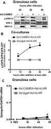
The selective activation of ERK1/2 in granulosa cells provokes the trans-phosphorylation of the EGFR in test cells in the absence of hCG stimulation. A, Primary cultures of granulosa cells were infected with Ad-hLHR and Ad-βgal alone or Ad-hLHR and Ad-CAMEK as indicated. Phosphorylated ERK1/2, total ERK2, and total MEK1/2 were measured using Western blots at different times after infection. The blot shown is representative of four experiments with similar results. B, Test cells expressing the EGFR-GFP were cocultured with primary granulosa cells expressing the hLHR and βgal (white symbols) or the hLHR and CAMEK (black symbols) at time zero. The phosphorylated and total EGFR-GFP was measured at different times after the cocultures were initiated. Note that the three time points shown in this panel correspond to the same time points shown in A and C for the individual cultures of granulosa cells. Each point is the mean ± sem of three independent experiments. Asterisks denote significant differences between the cells expressing CAMEK or βgal at a given time point (t test, P < 0.05). C, Primary cultures of granulosa cells were infected with Ad-hLHR and Ad-bgal (white symbols) or Ad-hLHR and Ad-CAMEK (black symbols) as indicated. Epiregulin and GAPDH mRNA were quantitated using RT followed by real-time PCR as described in Materials and Methods. Each point is the mean ± sem of five to six independent experiments. Asterisks denote significant differences between the cells expressing CAMEK or βgal at a given time point (t test, P < 0.05).
Figure 5
Figure 5
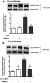
The hCG-induced trans-phosphorylation of the EGFR in test cells is blocked by an epiregulin antibody. Cocultures of test cells expressing the EGFR-GFP and primary immature granulosa cells expressing the hLHR were incubated without or with neutralizing antibodies (12.5 μg/ml) to epiregulin (A) or TGFα (B) for 15 min as indicated. Buffer (white bars) or 100 ng/ml hCG (black bars) was then added, and the incubation was continued for 9 h before measuring the phosphorylated and total EGFR-GFP. The blots shown are results of a representative experiment, whereas the graphs show the average ± sem of three independent experiments. Means with different letters are significantly different from each other (P < 0.05, ANOVA).
Figure 6
Figure 6
The trans-phosphorylation of the EGFR in test cells provoked by the selective phosphorylation of ERK1/2 in granulosa cells is blocked by an epiregulin antibody. Test cells expressing the EGFR-GFP were cocultured with primary granulosa cells expressing βgal (white bars) or CAMEK (black bars). Buffer or an antibody to epiregulin (12.5 μg/ml) was added as indicated at the beginning of the coculture and then again 8 h later. The phosphorylated EGFR-GFP and total EGFR-GFP were measured 12 h after the second addition of antibody. The blots shown are results of a representative experiment, whereas the graphs show the average ± sem of three independent experiments. Means with different letters are significantly different from each other (P < 0.05, ANOVA).
Figure 7
Figure 7
The hCG- or CAMEK-induced trans-phosphorylation of the EGFR in test cells is prevented by GM6001. A, Test cells expressing the EGFR-GFP were cocultured with primary granulosa cells expressing the hLHR. The cocultures were preincubated with GM6001 (20 μm) for 15 min before addition of buffer (white bars) or 100 ng/ml hCG (black bars). Phosphorylated and total EGFR-GFP was measured 9 h later. The blots shown are results of a representative experiment, whereas the graphs show the average ± sem of three independent experiments. Means with different letters are significantly different from each other (P < 0.05, ANOVA). B, Test cells expressing the EGFR-GFP were cocultured with primary rat granulosa cells expressing βgal (white bars) or CAMEK (black bars). Dimethylsulfoxide or GM6001 (20 μm) was added at the beginning of the coculture period and then again 8 h later. The phosphorylated EGFR-GFP and total EGFR-GFP were measured 12 h after the second addition of GM6001. The blots shown are results of a representative experiment, whereas the graphs show the average ± sem of three independent experiments. Means with different letters are significantly different from each other (P < 0.05, ANOVA).

References

    1. Liang CG, Su YQ, Fan HY, Schatten H, Sun QY 2007 Mechanisms regulating oocyte meiotic resumption: roles of mitogen-activated protein kinase. Mol Endocrinol 21:2037–2055 - PubMed
    1. Seger R, Hanoch T, Rosenberg R, Dantes A, Merz WE, Strauss III JF, Amsterdam A 2001 The ERK signaling cascade inhibits gonadotropin-stimulated steroidogenesis. J Biol Chem 276:13957–13964 - PubMed
    1. Andric N, Ascoli M 2006 A delayed, gonadotropin-dependent and growth-factor mediated activation of the ERK1/2 cascade negatively regulates aromatase expression in granulosa cells. Mol Endocrinol 20:3308–3320 - PMC - PubMed
    1. Zeleznik AJ, Saxena D, Little-Ihrig L 2003 Protein kinase B is obligatory for follicle-stimulating hormone-induced granulosa cell differentiation. Endocrinology 144:3985–3994 - PubMed
    1. Su YQ, Nyegaard M, Overgaard MT, Qiao J, Giudice LC 2006 Participation of mitogen-activated protein kinase in luteinizing hormone-induced differential regulation of steroidogenesis and steroidogenic gene expression in mural and cumulus granulosa cells of mouse preovulatory follicles. Biol Reprod 75:859–867 - PubMed

Publication types