. 2008 Jul 29;105(30):10559-64.
doi: 10.1073/pnas.0801674105. Epub 2008 Jul 24.

Native 3D intermediates of membrane fusion in herpes simplex virus 1 entry

Affiliations

Native 3D intermediates of membrane fusion in herpes simplex virus 1 entry

Ulrike E Maurer et al. Proc Natl Acad Sci U S A. .

Abstract

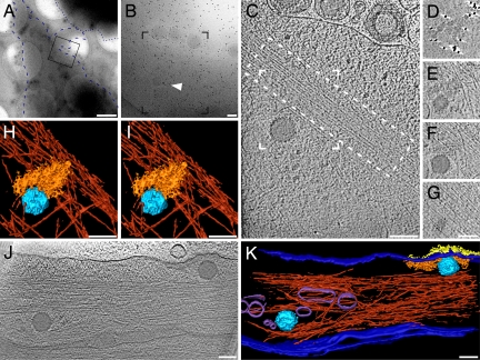
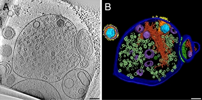
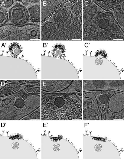
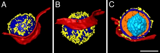
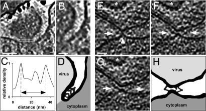
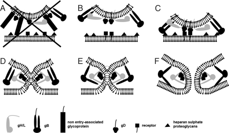
The concerted action of four viral glycoproteins and at least one cellular receptor is required to catalyze herpes simplex virus 1 entry into host cells either by fusion at the plasma membrane or intracellularly after internalization by endocytosis. Here, we applied cryo electron tomography to capture 3D intermediates from Herpes simplex virus 1 fusion at the plasma membrane in their native environment by using two model systems: adherent cells and synaptosomes. The fusion process was delineated as a series of structurally different steps. The incoming capsid separated from the tegument and was closely surrounded by the cortical cytoskeleton. After entry, the viral membrane curvature changed concomitantly with a reorganization of the envelope glycoprotein spikes. Individual glycoprotein complexes in transitional conformations during pore formation and dilation revealed the complex viral fusion mechanism in action. Snapshots of the fusion intermediates provide unprecedented details concerning the overall structural changes occurring during herpesvirus entry. Moreover, our data suggest that there are two functional "poles" of the asymmetric herpesvirion: one related to cell entry, and the other formed during virus assembly.


Conflict of interest statement

The authors declare no conflict of interest.

Figures

Fig. 1.
Fig. 1.
HSV-1 enters PtK2 cells by fusion at the plasma membrane. (A) Low-magnification electron micrograph of a PtK2 cell grown on a grid and inoculated with HSV-1 virions for 5 min at 37°C after virus binding on ice for 2 h and pretreatment with nocodazole. Whereas the central cellular areas were too thick for imaging and appear black, the cell periphery was sufficiently thin to allow tomographic imaging. The cell outline is accentuated by a blue, dashed line, a neighboring cell by a dotted blue line. The frame marks the area of a higher magnification projection image shown in B. Some virions are visible along the plasma membrane; the arrowhead highlights a capsid hardly recognizable in the projection image. Black dots are colloidal gold markers used for alignment of the tilt series. (C) A 3.2-nm-thick slice of the tomographic reconstruction of the boxed area in B reveals the cytosolic capsid and intracellular structures like macromolecular assemblies and actin filaments (dashed box). (D–G) Four different 3.2-nm-thick slices of the partial, denoised volume containing the capsid. (D) In a slice just above the plasma membrane, glycoprotein spikes cut in the stalk region appear as dots. (E) Directly underneath the plasma membrane, tegument protein density and the upper end of the capsid are visible. (F) An actin bundle neighbors the viral capsid and occupies the area underneath the capsid (G). (H–I) Stereoview of the surface rendering from the volume boxed in C. A dense actin network (red) underlies the incoming capsid (light blue) and the tegument (orange). The cell membrane, the glycoproteins, and macromolecular complexes are not displayed. (J) An 8.1-nm-thick slice from a tomogram of a PtK2 cell inoculated for 2 min in the absence of nocodazole. Two capsids of recently entered virions are visible. The one on the left entered from the top, and the right one from the side at the area where the glycoprotein spikes emerge from the membrane. (K) Surface rendering of the tomogram presented in J: capsid (light blue), tegument (orange), glycoproteins (yellow), cell membrane/viral membrane (dark blue), actin (dark red; upper part cut away), and cellular vesicles (purple). (Scale bars: A, 1 μm; and B–K, 100 nm.)
Fig. 2.
Fig. 2.
HSV-1 enters the presynaptic part of synaptosomes. (A) A 16-nm-thick slice of a tomogram from a synaptosome inoculated with HSV-1 for 60 min at 25°C. A capsid is localized inside the presynaptic element on the left characterized by abundant synaptic vesicles. A smaller postsynaptic fraction (right) is still attached via adhesion molecules to the synaptic cleft. Glycoprotein spikes are visible on the outer phase; tegument proteins correspond to the local densities near the cytoplasmic phase of the plasma membrane. Note structural changes between the entire virion (upper left corner) and the one that had entered the synaptosome. (B) Surface rendering of one virion and the synaptosome from the tomogram in A: capsid (light blue), tegument (orange), glycoproteins (yellow), cell membrane/viral membrane (dark blue), actin (dark red), vesicles (purple), synaptic vesicles (only partially segmented—metallic green), synaptic cleft (light green), and postsynaptic density (green). (Scale bars, 100 nm.)
Fig. 3.
Fig. 3.
Structural dynamics of HSV-1 during entry into synaptosomes. Different steps during HSV-1 entry into synaptosomes, for details refer to text. (A–F) Slices of 2.7 to 12.8 nm thickness of various tomographic reconstructions from HSV-1 entry intermediates; (A′ and F′) represent the corresponding schematic drawings. (A and A′) The virus attached to the plasma membrane by interaction of specific glycoproteins with cellular receptors. (B and B′) Fusion pore formation close to the capsid proximal side of the asymmetric virion (see Movie S3 and Fig. 4 for 3D representation). A part of the carbon film is present in the upper left. (C and C′) The capsid entered the cell, and the viral membrane was integrated into the cellular membrane while keeping its curvature, being most prominent in patches studded with glycoprotein spikes. (D and D′) The membrane was partly bent, and most of the tegument remained at the entry site. (E and E′) Glycoproteins were spread along the plasma membrane. (F and F′) The membrane curvature returned to a more flattened, regular organization, and the glycoproteins were removed from the surface. (Scale bars, 100 nm.)
Fig. 4.
Fig. 4.
Detailed analysis of a fusion pore. (A–C) Surface renderings of the volume from Fig. 3B in different orientations (see Movie S3 for complete tomogram). (A) Rendering of the virus with parts of the synaptosomal plasma membrane in the same orientation as in the tomographic slice shown in Fig. 3B and Movie S3. (B) The virus was rotated vertically for ≈60° to allow a better view into the fusion pore. (C) Cut-away view of all features apart from the capsid; the orientation was changed to provide a perpendicular view of the pore. Colors used are light blue (capsid), orange (tegument), yellow (glyco proteins), dark blue (viral membrane), and red (cell membrane). Cellular elements of the synaptosome were excluded from rendering for clarity. (Scale bar, 100 nm.)
Fig. 5.
Fig. 5.
Glycoprotein bridges are formed prior and next to the fusion pore. (A) A 2.7-nm-thick slice from the volume presented in Fig. 4. Continuous V- and Y-shaped structures are localized between the two membranes. (B) Zoom into the area marked by box corners in A. (C) Line plot of pixel intensities from the pore cross-section (see rectangle in A). The pore diameter of this entry intermediate was ≈25 nm as measured between the maxima for the membrane. The gray values within the pore are higher than those of the surrounding vitreous ice. (D) Schematic drawing of membrane and protein structures from A with emphasis on connective densities and membranes. (E–G) Subsequent 2.7-nm-thick slices from a tomogram of an inoculated, nocodazole-treated Vero cell. The capsid of the virion is marked by a star. The arrowhead (E) highlights the contact between the plasma membrane and the viral envelope. The arrows in (E–G) point to prominent protein complexes between the two membranes and close to the contact site. (H) Schematic drawing of the connecting densities and membrane structures in F and G. (Scale bars; A, 40 nm; B and E, 20 nm.)
Fig. 6.
Fig. 6.
Schematic model for the fusion mechanism based on the identified intermediates. (A) The bulky glycoprotein spikes in close proximity to gD, which are not involved in cell entry, act as spacers. Thus, gD cannot bind to its specific receptor, and the fusion process is not initiated. (B) Fusion can only be initiated if the contact of the entry-associated glycoprotein gD with its cellular receptor is not sterically hindered. (C) Binding of gD to its receptor might be paralleled or preceded by nonessential interactions of gB with heparan sulfate proteoglycans. (D) After receptor binding, gD undergoes a conformational change and interacts with gH/gL. This induces gH/gL to also change conformation and to flip out one segment that binds to the plasma membrane. This connects the two membranes, brings them close together and enables lipid mixing (E). Glycoprotein B is recruited to the gD/gH/gL complex by the conformational switch of gH/gL and binds to the plasma membrane (F). The fusion pore forms stabilized by gB.

References

    1. Grünewald K, et al. Three-dimensional structure of Herpes simplex virus from cryo-electron tomography. Science. 2003;302:1396–1398. - PubMed
    1. Spear PG. Herpes simplex virus: Receptors and ligands for cell entry. Cell Microbiol. 2004;6:401–410. - PubMed
    1. Nicola AV, Hou J, Major EO, Straus SE. Herpes simplex virus type 1 enters human epidermal keratinocytes, but not neurons, via a pH-dependent endocytic pathway. J Virol. 2005;79:7609–7616. - PMC - PubMed
    1. Rey FA. Molecular gymnastics at the herpesvirus surface. EMBO Rep. 2006;7:1000–1005. - PMC - PubMed
    1. Heldwein EE, Krummenacher C. Entry of herpesviruses into mammalian cells. Cell Mol Life Sci. 2008;65:1653–1668. - PMC - PubMed

Publication types