. 2008 Jul 29;105(30):10571-6.
doi: 10.1073/pnas.0802039105. Epub 2008 Jul 24.

A positive-feedback mechanism promotes reovirus particle conversion to the intermediate associated with membrane penetration

Affiliations

A positive-feedback mechanism promotes reovirus particle conversion to the intermediate associated with membrane penetration

Melina A Agosto et al. Proc Natl Acad Sci U S A. .

Abstract

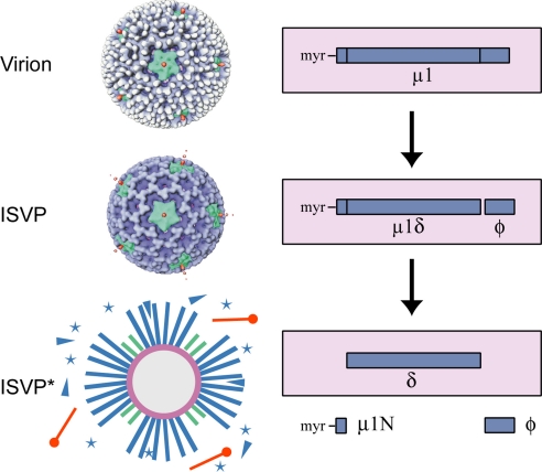
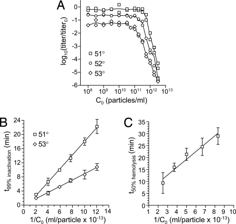
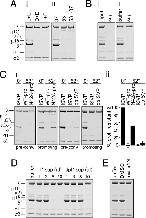
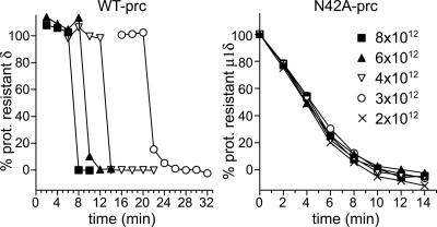
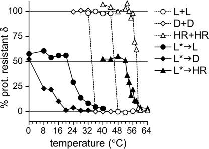
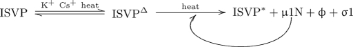
Membrane penetration by reovirus is associated with conversion of a metastable intermediate, the ISVP, to a further-disassembled particle, the ISVP*. Factors that promote this conversion in cells are poorly understood. Here, we report the in vitro characterization of a positive-feedback mechanism for promoting ISVP* conversion. At high particle concentration, conversion approximated second-order kinetics, and products of the reaction operated in trans to promote the conversion of target ISVPs. Pore-forming peptide mu1N, which is released from particles during conversion, was sufficient for promoting activity. A mutant that does not undergo mu1N release failed to exhibit second-order conversion kinetics and also failed to promote conversion of wild-type target ISVPs. Susceptibility of target ISVPs to promotion in trans was temperature dependent and correlated with target stability, suggesting that capsid dynamics are required to expose the interacting epitope. A positive-feedback mechanism of promoting escape from the metastable intermediate has not been reported for other viruses but represents a generalizable device for sensing a confined volume, such as that encountered during cell entry.


Conflict of interest statement

The authors declare no conflict of interest.

Figures

Fig. 1.
Fig. 1.
Reovirus particles and μ1 protein. (Left) Electron cryomicroscopy reconstructions of virions and ISVPs (3) and a diagram of ISVP*s. White, σ3; blue, μ1; green, λ2; orange, σ1; stars, released μ1N; triangles, released φ. (Right) Diagram of μ1 status in each particle type. Species inside the box are associated with particles; those outside the box are released. At least 50% of the μ1 copies in virions and ISVPs consist of full-length μ1 and μ1δ, respectively (containing uncleaved μ1), although the exact amount of cleavage is unknown (18, 19). Because standard disruption conditions for SDS/PAGE lead to μ1N cleavage (18), the main μ1 species seen in gels of ISVPs in this study is δ. μ1δ and μ1C (resulting from cleavage of μ1N only) are seen as minor species.
Fig. 2.
Fig. 2.
ISVP* conversion approximates a second-order reaction at high particle concentrations. (A) ISVPs at the indicated C0 were subjected to heat treatment at 51, 52, or 53°C for 15 min, then titered by plaque assay. Titer loss is expressed as log10 change relative to a 0°C sample. Points from duplicate experiments are shown with a line connecting mean values. (B) Short time courses bracketing the time required for a 99% reduction in titer (t99%) were performed over a range of C0. The expected linear relationship for a second-order reaction is plotted. Means ± SD for three or four determinations are shown with a linear-regression fit. (C) Hemolysis reactions containing ISVPs at the indicated C0 were performed at 37°C in buffer containing Cs+, which facilitates conversion. The expected linear relationship between time to half-maximal hemolysis (t50%) and concentration is plotted. Means ± SD for four or five determinations are shown with a linear-regression fit line. Examples of time courses used to generate the points in B and C are shown in Fig. S1.
Fig. 3.
Fig. 3.
Released μ1N is sufficient for promoting activity. (A) (i) ISVPs of T1L (L+L) or T3D (D+D), or a mixture of equal parts of both (L+D), were incubated at 37°C for 60 min. ISVP* conversion was assayed by TRY sensitivity of the particle-associated μ1 species (μ1C, μ1δ, and δ). (ii) T1L ISVPs were incubated at 37°C for 65 min (lane 1); at 53°C for 5 min (lane 2); or preconverted at 53°C for 5 min, chilled and supplemented with an equal part of fresh ISVPs, and incubated at 37°C for 60 additional min (lane 3). ISVP* conversion was assayed by TRY sensitivity. (B) ISVPs were preconverted at 52°C and centrifuged to pellet particles. (i) Equivalent amounts of input particles and supernatant, demonstrating efficient particle clearance (μ1N and φ are too small to be resolved on this gel). (ii) 10 μl of buffer or supernatant were added to ISVPs, and reactions were incubated at 37°C for 30 min followed by the TRY-sensitivity assay. (C) (i) WT-pRCs, N42A-pRCs, ISVPs, and dpISVPs were preconverted at 52°C. Preconversion of a duplicate sample was confirmed by the TRY-sensitivity assay (“pre-conv.”). Preconverted particles were chilled and supplemented with fresh WT ISVPs, incubated at 37°C for 30 min, and assayed by TRY sensitivity (“promoting”). Samples containing ISVPs incubated at 0°C, instead of 52°C, were analyzed in parallel. (ii) Quantitation of three or more experiments such as shown in i. Two different preparations of each particle type were used. The amount of δ remaining after the TRY-sensitivity assay is expressed relative to the amount in samples containing only unconverted ISVPs (0° lanes in i). The 50% protease-resistant δ in the N42A-pRC lane reflects preconversion of the N42A-pRCs but no promotion of the target ISVPs, which are present in equal numbers. Error bars indicate SD. (D) ISVPs or dpISVPs were preconverted at 52°C and centrifuged to pellet particles. The indicated amounts of supernatant were added to fresh WT ISVPs, and reactions were incubated at 37°C for 30 min followed by TRY-sensitivity assay. (E) ISVPs were incubated with 50 μg/ml synthetic μ1N peptide or vehicle control (1% DMSO) at 37°C for 60 min followed by TRY-sensitivity assay. All reactions contained a total final ISVP concentration of 5 × 10¹² particles per milliliter.
Fig. 4.
Fig. 4.
μ1N cleavage is required for concentration-dependent ISVP* conversion. WT- or N42A-pRCs at the indicated concentrations were incubated at 37°C in buffer containing Cs+, which facilitates conversion. At each time point, a sample was assayed for TRY sensitivity, and the δ remaining after TRY digestion (or μ1δ, in the case of N42A-pRCs) was quantitated. Representative experiments are shown. Gels used for quantitation are shown in Fig. S2.
Fig. 5.
Fig. 5.
Susceptibility of target ISVPs to promotion in trans is temperature dependent. Aliquots of WT T1L ISVPs were preconverted at 52°C, chilled on ice, and supplemented with an equal part of T1L ISVPs (L*→L), T3D ISVPs (L*→D), or ISVPs of the heat-resistant mutant IL62–3 (L*→HR). Alternatively, reactions contained only T1L ISVPs (L+L), T3D ISVPs (D+D), or IL62–3 ISVPs (HR+HR), without any preconverted particles. Reactions were incubated at the indicated temperature and then assayed for TRY sensitivity, and the δ remaining after TRY digestion was quantitated. For samples containing preconverted ISVPs, 50% protease-resistant δ indicates no promoting activity because the preconverted ISVPs and target ISVPs are present in equal parts. Each point represents the mean of two or three determinations.
Fig. 6.
Fig. 6.
Model of ISVP* conversion. See Discussion for details.

References

    1. Nibert ML, Schiff LA, Fields BN. Mammalian reoviruses contain a myristoylated structural protein. J Virol. 1991;65:1960–1967. - PMC - PubMed
    1. Tillotson L, Shatkin AJ. Reovirus polypeptide σ3 and N-terminal myristoylation of polypeptide μ1 are required for site-specific cleavage to μ1C in transfected cells. J Virol. 1992;66:2180–2186. - PMC - PubMed
    1. Dryden KA, et al. Early steps in reovirus infection are associated with dramatic changes in supramolecular structure and protein conformation: Analysis of virions and subviral particles by cryoelectron microscopy and image reconstruction. J Cell Biol. 1993;122:1023–1041. - PMC - PubMed
    1. Liemann S, Chandran K, Baker TS, Nibert ML, Harrison SC. Structure of the reovirus membrane-penetration protein, μ1, in a complex with is protector protein, σ3. Cell. 2002;108:283–295. - PMC - PubMed
    1. Furlong DB, Nibert ML, Fields BN. Sigma 1 protein of mammalian reoviruses extends from the surfaces of viral particles. J Virol. 1988;62:246–256. - PMC - PubMed

Publication types

LinkOut - more resources