. 2007 Nov;2(11):718-24.
doi: 10.1038/nnano.2007.344. Epub 2007 Oct 28.

Sequence-specific detection of individual DNA polymerase complexes in real time using a nanopore

Sequence-specific detection of individual DNA polymerase complexes in real time using a nanopore

Seico Benner et al. Nat Nanotechnol. 2007 Nov.

Abstract

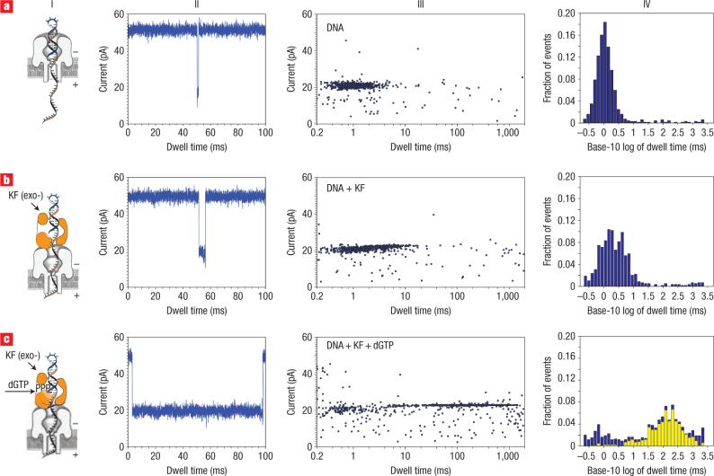
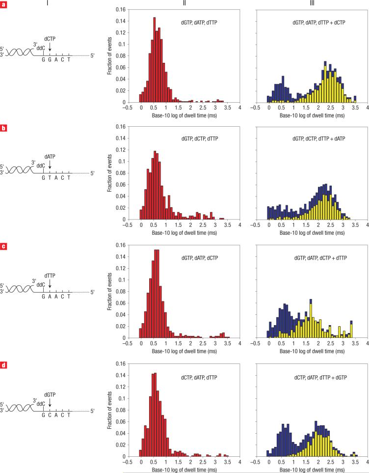
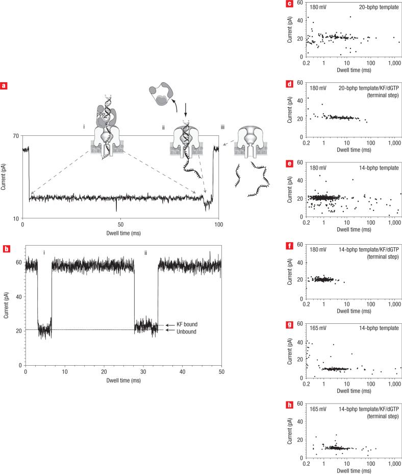
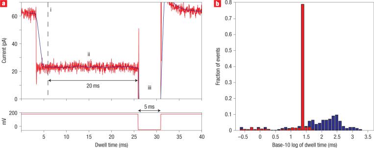
Nanoscale pores have potential to be used as biosensors and are an established tool for analysing the structure and composition of single DNA or RNA molecules. Recently, nanopores have been used to measure the binding of enzymes to their DNA substrates. In this technique, a polynucleotide bound to an enzyme is drawn into the nanopore by an applied voltage. The force exerted on the charged backbone of the polynucleotide by the electric field is used to examine the enzyme-polynucleotide interactions. Here we show that a nanopore sensor can accurately identify DNA templates bound in the catalytic site of individual DNA polymerase molecules. Discrimination among unbound DNA, binary DNA/polymerase complexes, and ternary DNA/polymerase/deoxynucleotide triphosphate complexes was achieved in real time using finite state machine logic. This technique is applicable to numerous enzymes that bind or modify DNA or RNA including exonucleases, kinases and other polymerases.


Figures

Figure 1
Figure 1. Detection of DNA translocation events with the nanopore device
a, Schematic of the nanopore device. Voltage is applied across a single α-haemolysin channel inserted in a ∼25-μm-diameter lipid bilayer (trans-side positive). Current through the nanopore is carried by K+ and Cl ions (0.3 M bulk phase concentration). b, Current trace for the primer/template DNA being captured, and then translocated through the nanopore under an applied voltage (bottom). An illustration of the events that yield the three sections of the current trace is shown over the trace. Io is the open channel current, I is the average current level for the blockade event, and tD is the translocation dwell time.
Figure 2
Figure 2. Distinguishing DNA, DNA/KF complexes or DNA/KF/dNTP complexes in the nanopore device
a, Translocation, through the nanopore, of DNA alone (14-bp hairpin with a 36-nucleotide 5′ overhang and 2′–3′ dideoxycytidine terminus; template base at n = 0 is C). b,c, Translocation of the 14-bphp from complexes with KF (b) or from complexes with KF and dGTP (c). For each of rows a–c are presented a diagram of the nanopore with the associated complex (column I), a current trace (column II) and a dwell time event plot (column III). In column IV, probability histograms of the base-10 logarithm of dwell time data are shown in blue. Close examination of the event plot in c, column III, reveals that most long dwell time events are within 22 to 24 pA. A yellow subset histogram for the events within 22−24 pA is overlaid on probability histogram c, revealing that the chosen range is dominated by long dwell time events. The number of events, mean and IQR values for probability histograms a and b, and subset histogram c, are reported in Table 1.
Figure 3
Figure 3. Detection of correct dNTP binding to the KF/primer-template complex
Column I depicts four primer/template hybrids, which differ only in the template base (position n = 0). The primer has a 3′ ddC terminus, and the correct incoming dNTP, complementary to n = 0, is indicated. a–d, Translocation event dwell time histograms when n = 0 is G (a), T (b), A (c) and C (d), respectively. Column II shows probability histograms of event dwell times for primer/template pairs in the presence of KF and all three incorrect dNTPs (in red). Column III shows probability histograms after the fourth complementary dNTP was added (in blue). Most long dwell time events are within the range of −35 to −32 pA (a,b,d) and −41 to −37 pA (c). Subset histograms for the events within these ranges (in yellow) are overlaid on the probability histograms. The number of events, mean and IQR values for probability histograms (II) and subset histograms (III) are reported in Table 1. Because the histograms in this figure are based on data from multiple experiments, blockade events are reported relative to a 0 pA open channel current. Consequently, the reported blockade amplitudes fall within the −50 to −20 pA range.
Figure 4
Figure 4. Proposed mechanism for voltage-facilitated dissociation of DNA from KF/DNA or KF/DNA/dNTP complexes
a, Representative trace at 180 mV showing the current levels associated with nanopore capture of the ternary complex, and illustration of the molecular events proposed to occur at each step. i, The long 24 pA blockade arises from the initial capture of the ternary complex, with the duplex DNA held on top the pore vestibule because of association with the KF. ii, The shorter 20 pA terminal step occurs upon KF dissociation, when duplex DNA is pulled into the nanopore vestibule. iii, Finally, primer and template strands are dissociated, translocating through the nanopore as single-stranded molecules and resulting in a return to the open channel current. b, Current trace from an experiment with DNA and KF in the nanopore chamber, showing two translocation events. i, Trace shows the 20 pA signature characteristic of events in the presence of DNA only. ii, Trace is characteristic of events that only occur in the presence of both KF and DNA (without the correct dNTP). c–h, Two-dimensional plots show event clusters for 20-bp DNA hairpin translocation at 180 mV (c); 20-bp DNA hairpin/KF/dGTP complex terminal step at 180 mV (d); 14-bp DNA hairpin translocation at 180 mV (e); 14-bp DNA hairpin/KF/dGTP complex terminal step at 180 mV (f); 14-bp DNA hairpin translocation at 165 mV (g); 14-bp DNA hairpin/KF/dGTP complex terminal step at 165 mV (h).
Figure 5
Figure 5. Recognition and control of DNA complexes in real time using FPGA
a, A representative ternary complex event under FPGA control. The FPGA samples the ionic current every 5.3 μs and computes a windowed mean amplitude (solid dark line) based on the previous 1.5 ms of signal. Every 0.4 ms, it tests whether or not the mean is within 24 ± 2.8 pA. i, If the mean remains within this range for four consecutive tests, the FSM logic diagnoses the blockade as a KF binding event. The total delay for diagnosis of a KF binding event is 2.7 ms (1.5 ms for the windowed mean to enter the 24 ± 2.8 pA range, plus 1.2 ms for three consecutive subsequent tests). ii, Upon diagnosis of a KF binding event, the FPGA continues to monitor the windowed mean. If the mean remains in the 24 ± 2.8 pA range for 20 ms, the FSM logic diagnoses the blockade as resulting from a ternary complex. The 20 ms cutoff was used because 60% of events are longer than 20 ms in the presence of the correct dNTP, but only 2% of events are longer than 20 ms in the 24 ± 2.8 pA range in the absence of the correct dNTP. iii, Upon diagnosis that a ternary complex is in the pore, the FPGA reverses the voltage to −50 mV for 5 ms, ejecting the complex from the pore. The 180 mV capture voltage is then restored. For each event in which KF binding is not diagnosed, or in which KF binding is diagnosed but the mean leaves the 24 ± 2.8 pA range before 20 ms, the voltage is kept at 180 mV. b, Dwell time probability histograms for 24 ± 2.8 pA events with FPGA control (527 total events, in red) and without FPGA control (155 total events, in blue).

References

    1. Deamer DW, Branton D. Characterization of nucleic acids by nanopore analysis. Acc. Chem. Res. 2002;35:817–825. - PubMed
    1. Dekker C. Solid-state nanopores. Nature Nanotechnol. 2007;2:209–215. - PubMed
    1. Siwy Z, et al. Protein biosensors based on biofunctionalized conical gold nanotubes. J. Am. Chem. Soc. 2005;127:5000–5001. - PubMed
    1. Hornblower B, et al. Single-molecule analysis of DNA–protein complexes using nanopores. Nature Methods. 2007;4:315–317. - PubMed
    1. Zhao Q, et al. Detecting SNPs using a synthetic nanopore. Nano Lett. 2007;7:1680–1685. - PMC - PubMed

Publication types