. 2008 Jul;3(7):418-22.
doi: 10.1038/nnano.2008.164. Epub 2008 Jun 22.

Self-assembled DNA nanostructures for distance-dependent multivalent ligand-protein binding

Self-assembled DNA nanostructures for distance-dependent multivalent ligand-protein binding

Sherri Rinker et al. Nat Nanotechnol. 2008 Jul.

Abstract

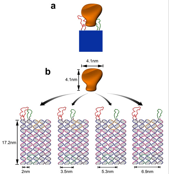
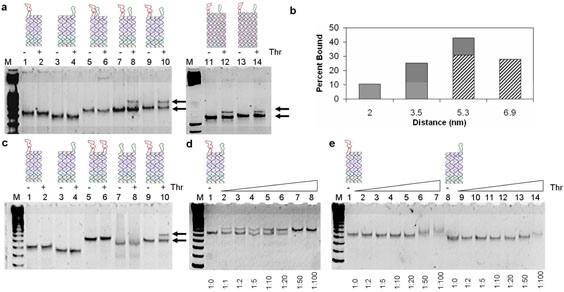
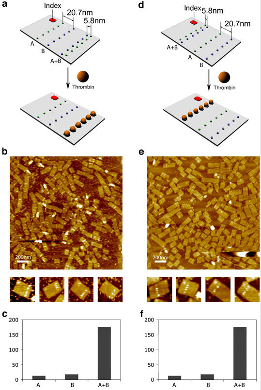
An important goal of nanotechnology is to assemble multiple molecules while controlling the spacing between them. Of particular interest is the phenomenon of multivalency, which is characterized by simultaneous binding of multiple ligands on one biological entity to multiple receptors on another. Various approaches have been developed to engineer multivalency by linking multiple ligands together. However, the effects of well-controlled inter-ligand distances on multivalency are less well understood. Recent progress in self-assembling DNA nanostructures with spatial and sequence addressability has made deterministic positioning of different molecular species possible. Here we show that distance-dependent multivalent binding effects can be systematically investigated by incorporating multiple-affinity ligands into DNA nanostructures with precise nanometre spatial control. Using atomic force microscopy, we demonstrate direct visualization of high-affinity bivalent ligands being used as pincers to capture and display protein molecules on a nanoarray. These results illustrate the potential of using designer DNA nanoscaffolds to engineer more complex and interactive biomolecular networks.


Figures

Figure 1
Figure 1. Schematics of the self-assembled divalent aptamers on DNA tile for protein binding
a, A cartoon showing a rigid DNA tile (blue) that can spatially separate two ligands (red and green) at a controlled distance with each ligand attaches to a different part of the target molecule (orange) for bivalent binding. b, 5-helix-bundle (5HB) DNA structure with apt-A (red) and apt-B (green) protruding out of the tile helices and being separated at a distance of 2, 3.5, 5.3, and 6.9 nm. The aptamer sequences were incorporated into the closed loops extending out of the ends of the helices. Apt-A is fixed on helix 1 and apt-B is moved between helix 2 and helix 5, generating the varying inter-aptamer distances but keeping the relative orientations constant. Numbering of the helices in the tile is read from left to right.
Figure 2
Figure 2. Gel-mobility shift assays
a, Nondenaturing (8% polyacrylamide) PAGE image of aptamers linked on the 4HB and 5HB tiles. Lane M corresponds to a 25bp DNA marker. Lanes 1 to 14 correspond to 4HB-A1, 4HB-B4, 4HB-A1- B2 (2 nm), 4HB-A1-B3 (3.5 nm), 4HB-A1-B4 (5.3 nm), 5HB-A1-B4 (5.3nm), and 5HB-A1-B5 (6.9 nm), each 20 nM without (−) and with (+) thrombin (40 nM), respectively. The lower band in each lane represents the unbound tiles and the upper band in lane 6, 8, 10, 12 and 14 represents the tile/thrombin complex. b, The dependence of protein binding on the inter-aptamer distance. Here 20 nM 4HB or 5HB tiles containing both apt-A and apt-B with varying distances were incubated with 40 nM thrombin before they were loaded into the gel. The 16 percentages of bound DNA tiles were estimated from the relative intensity of the bands in the gel image shown in panel a. The filled bar and striped bars are data from the 4-HB and 5-HB tiles respectively. c, The binding of thrombin to the 4HB tile-based aptamer structures carrying one aptamer or two of the same or different aptamers. All tiles with two aptamers have the same inter-aptamer distance at 5.3 nm. Lane M corresponds to a 25 bp DNA marker. Lanes 1, to 10 correspond to 4HB-A1, 4HB-B4, 4HB-A1-A4, 4HB-B1-B4, and 4HB-A1-B4 without (−) and with (+) thrombin (40 nM). An upper band is only observed in lane 10 with 4HB-A1-B4 (same as lane 10 in panel a). d, Titration experiment showing more DNA tiles (4HB-A1-B4, 1 nM) are bound to the protein with increasing concentrations of thrombin. Lane M corresponds to a 25 bp DNA ladder. From this gel a 10 nM apparent KD is estimated for the bivalent binding of thrombin to the two aptamers linked by a DNA tile at a 5.3 nm distance. e, Titration experiment for 4HB-A1 (lanes 1-7) and 4HB-B4 (lanes 8-14). Lane M is a 25 pb DNA ladder. From this gel, the apparent KD for the two individual aptamers on the 4HB tile are estimated to be 20-50 nM and > 50 nM, respectively.
Figure 3
Figure 3. Evaluation of bivalent binding by AFM
a, d, schematic drawings of two rectangular-shaped DNA origami tiles containing two lines of apt-A (green dots) and two lines of apt-B thrombin (blue dots). The neighboring lines of apt-A and apt-B are ~ 20.7 nm (marked line A and line B) and ~ 5.8 nm (line A+B) apart. We call the closely spaced lines the dual-aptamer line. An index is also included helping to verify the positions of the lines in the AFM images. b, e, AFM height 17 images of the DNA origami tiles (10nM) with 60 nM thrombin, corresponding to a and d, respectively. Zoom-in images are 150nm × 150nm. c, f, Charts of numbers of proteins binding on each aptamer line (observed from 60 arrays corresponding to a and d respectively). Bar A represents the number of proteins on the line of apt-A, bar B represents the number of proteins on the line of apt-B, and bar A+B represents the number of proteins on the bivalent dual aptamer line.

References

    1. Mammen M, Choi SK, Whitesides GM. Polyvalent interactions in biological systems: implications for design and use of multivalent ligands and inhibitors. Angew. Chem. Int. Ed. 1998;37:2755–2794. - PubMed
    1. Di Giusto DA, King GC. Construction, stability, and activity of multivalent circular anticoagulant aptamers. J. Biol. Chem. 2004;279:46483–46489. - PubMed
    1. Fredriksson S, et al. Protein detection using proximity-dependent DNA ligation assays. Nat. Biotechnol. 2002;20:473–477. - PubMed
    1. Pei R, et al. Behavior of polycatalytic assemblies in a substrate-displaying matrix. J. Am. Chem. Soc. 2006;128:12693–12699. - PubMed
    1. Rothemund PWK, Papadakis N, Winfree E. Algorithmic self-assembly of DNA sierpinski triangles. PloS Biology. 2004;2:2041–2053. - PMC - PubMed

Publication types