. 2008 Aug;118(8):2771-84.
doi: 10.1172/JCI32709.

Cigarette smoke selectively enhances viral PAMP- and virus-induced pulmonary innate immune and remodeling responses in mice

Affiliations

Cigarette smoke selectively enhances viral PAMP- and virus-induced pulmonary innate immune and remodeling responses in mice

Min-Jong Kang et al. J Clin Invest. 2008 Aug.

Abstract

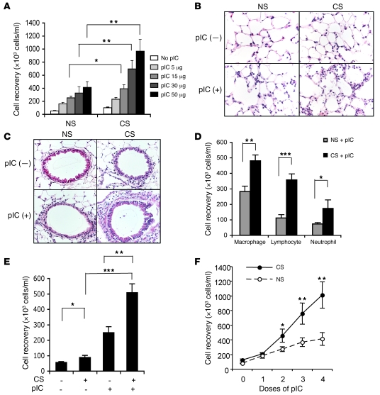
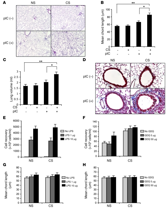
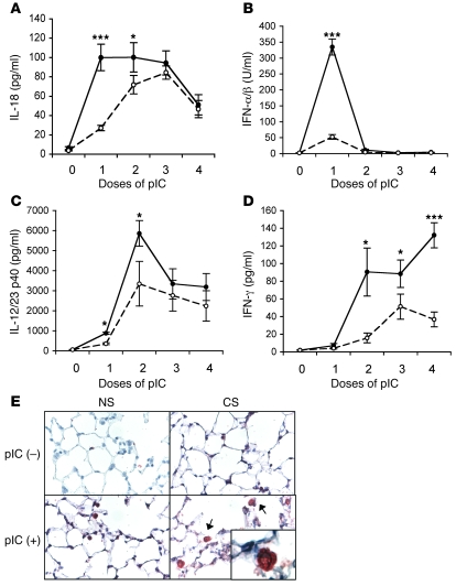
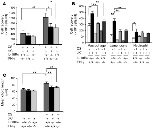
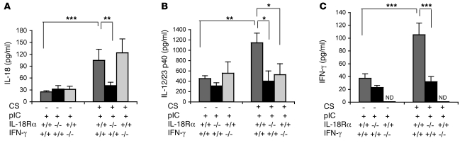
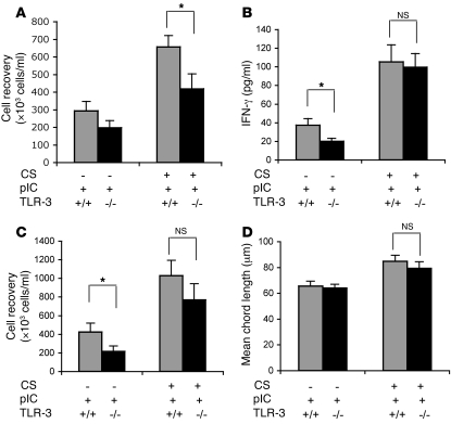
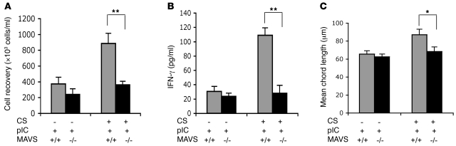
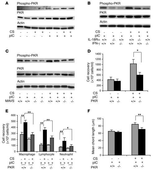
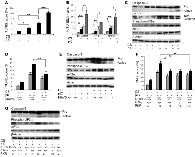
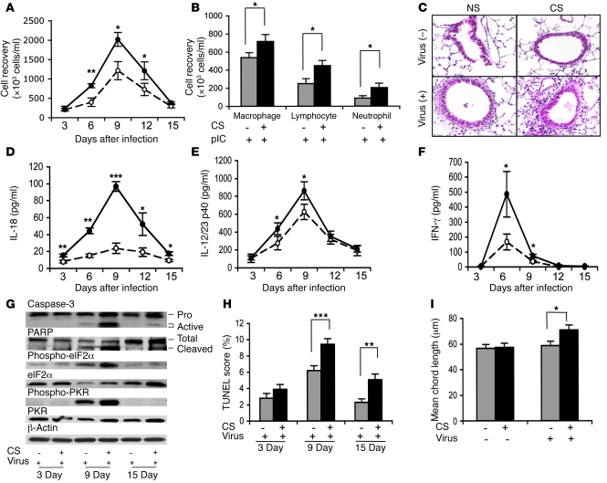
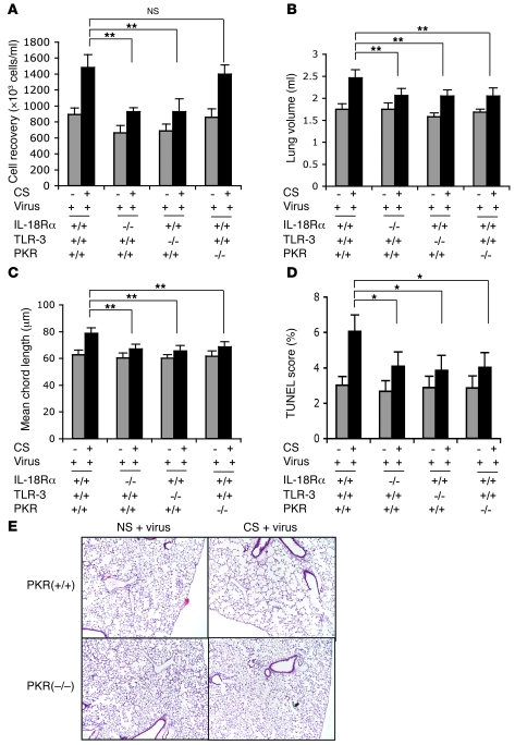
Viral infections have more severe consequences in patients who have been exposed to cigarette smoke (CS) than in those not exposed to CS. For example, in chronic obstructive pulmonary disease (COPD), viruses cause more severe disease exacerbation, heightened inflammation, and accelerated loss of lung function compared with other causes of disease exacerbation. Symptomatology and mortality in influenza-infected smokers is also enhanced. To test the hypothesis that these outcomes are caused by CS-induced alterations in innate immunity, we defined the effects of CS on pathogen-associated molecular pattern-induced (PAMP-induced) pulmonary inflammation and remodeling in mice. CS was found to enhance parenchymal and airway inflammation and apoptosis induced by the viral PAMP poly(I:C). CS and poly(I:C) also induced accelerated emphysema and airway fibrosis. The effects of a combination of CS and poly(I:C) were associated with early induction of type I IFN and IL-18, later induction of IL-12/IL-23 p40 and IFN-gamma, and the activation of double-stranded RNA-dependent protein kinase (PKR) and eukaryotic initiation factor-2alpha (eIF2alpha). Further analysis using mice lacking specific proteins indicated a role for TLR3-dependent and -independent pathways as well as a pathway or pathways that are dependent on mitochondrial antiviral signaling protein (MAVS), IL-18Ralpha, IFN-gamma, and PKR. Importantly, CS enhanced the effects of influenza but not other agonists of innate immunity in a similar fashion. These studies demonstrate that CS selectively augments the airway and alveolar inflammatory and remodeling responses induced in the murine lung by viral PAMPs and viruses.


Figures

Figure 1
Figure 1. Inflammatory effects of poly(I:C) in mice exposed to RA or CS.
C57BL/6J mice were exposed to CS or RA (NS, nonsmoking) for 2 weeks and then randomized to receive 4 doses of poly(I:C) [pIC+] or vehicle control [pIC–]. The effects of 4 doses of varying concentrations of poly(I:C) on BAL total cell recovery (A), 4 doses of poly(I:C) (50 μg) on parenchymal (B) and airway (C) inflammation, and BAL differential cell recovery (D) are illustrated. (E) Effects of 4 doses of poly(I:C) on BALB/c mice breathing RA (CS–) and mice exposed to CS (CS+) are illustrated. (F) Dose dependence of these inflammatory events in C57BL/6J mice. The values in parts A, D, E, and F represent the mean ± SEM of evaluations in a minimum of 5 mice. Parts B and C are representative of a minimum of 4 similar experiments. *P < 0.05; **P < 0.01; ***P < 0.001. Original magnification, ×20.
Figure 2
Figure 2. Effects of poly(I:C) and other innate immunity agonists on mice exposed to RA or CS.
Mice were exposed to CS or RA for 2 weeks and then randomized to receive 4 doses of poly(I:C) (50 μg), LPS (1–10 μg), GDQ (5–50 μg), or vehicle controls. The alterations in alveolar structure, alveolar chord length, and lung volume caused by poly(I:C) are noted in parts A, B, and C. The effects of these interventions on matrix accumulation were assessed with trichrome evaluations (D). The effects of LPS and GDQ on BAL inflammation are seen in E and F. The effects of LPS and GDQ on alveolar remodeling are illustrated in parts G and H, respectively. The values in B, C, and EH represent the mean ± SEM of evaluations in a minimum of 5 mice. Parts A and C are representative of a minimum of 4 similar experiments. *P < 0.05; **P < 0.01. Original magnification, ×4 (A); ×20 (D).
Figure 3
Figure 3. Effects of poly(I:C) on cytokines and IFNs.
Mice were exposed to CS or RA for 2 weeks and then randomized to receive poly(I:C) (50 μg) or vehicle control. The levels of BAL IL-18, IFN-α/β, IL-12/IL-23 p40, and IFN-γ in vehicle-treated mice breathing RA or CS were near or below the limits of detection of these assays. The effects of poly(I:C) in mice exposed to CS (filled circles) or RA (open circles) are illustrated (AD). The localization of IL-18 was accomplished using IHC (E). In the lungs from mice exposed to CS plus poly(I:C), selected IL-18–containing macrophages are highlighted with arrows, and a high-power image can be seen in the insert. The values in parts AD represent the mean ± SEM of evaluations in a minimum of 5 mice. Part E is representative of 3 similar experiments. *P < 0.05; ***P < 0.001. Original magnification, ×20 (E); ×100 (insert).
Figure 4
Figure 4. Roles of IL-18Rα and IFN-γ in the interaction of CS and poly(I:C).
WT (+/+) mice and mice with null mutations (–/–) of IL-18Rα or IFN-γ were exposed to CS or RA (CS–) for 2 weeks and then given 4 doses of poly(I:C) (50 μg) or vehicle control. BAL total cell recovery (A), BAL differential cell recovery (B), and emphysema (C) were evaluated. The noted values represent the mean ± SEM of evaluations in a minimum of 5 mice. *P < 0.05; **P < 0.01.
Figure 5
Figure 5. Roles of IL-18Rα and IFN-γ in CS plus poly(I:C)–induced cytokine stimulation.
WT (+/+) mice and mice with null mutations (–/–) of IL-18Rα or IFN-γ were exposed to CS or RA (CS–) for 2 weeks and then given 1 (A and B) or 4 doses (C) of poly(I:C) (50 μg) or vehicle control. BAL IL-18 (A), IL-12/IL-23 p40 (B), and IFN-γ (C) levels were quantitated. The noted values represent the mean ± SEM of evaluations in a minimum of 5 mice. *P < 0.05; **P < 0.01; ***P < 0.001. ND, none detected.
Figure 6
Figure 6. Roles of TLR3 in the interaction of CS and poly(I:C).
WT (+/+) mice and mice with null mutations (–/–) of TLR3 were exposed to CS or RA (CS–) for 2 weeks and then given 1 or 4 doses of poly(I:C) (50 μg) or vehicle control. BAL total cell recovery after a single dose of poly(I:C) is illustrated in A. The levels of BAL IFN-γ (B), BAL total cell recovery (C), and alveolar remodeling (D) after 4 doses of poly(I:C) were also evaluated. The noted values represent the mean ± SEM of evaluations in a minimum of 5 mice. *P < 0.05.
Figure 7
Figure 7. Roles of MAVS in the interaction of CS and poly(I:C).
WT (+/+) mice and mice with null mutations (–/–) of MAVS were exposed to CS or RA (CS–) for 2 weeks and then given 4 doses of poly(I:C) (50 μg) or vehicle control. BAL total cell recovery (A), IFN-γ production (B), and alveolar remodeling (C) were evaluated. The noted values represent the mean ± SEM of evaluations in a minimum of 5 mice. *P < 0.05; **P < 0.01.
Figure 8
Figure 8. Regulation and roles of PKR in the interaction of CS and poly(I:C).
Mice were exposed to CS or RA (CS–) for 2 weeks and then given 4 doses of poly(I:C) (50 μg) or vehicle control. (A) Effects of poly(I:C) on PKR phosphorylation. The roles of IL-18Rα and IFN-γ in this activation are seen in comparisons of the levels of PKR phosphorylation in WT (+/+) mice and mice with null (–/–) mutations of IL-18Rα or IFN-γ (B). The role or roles of MAVS in this activation are seen in comparisons of the levels of PKR phosphorylation in WT (+/+) mice and mice with null (–/–) mutations of MAVS (C). The roles of PKR in poly(I:C)-induced total (D) and differential (E) BAL cell recovery and emphysema (F) are also illustrated. The values in parts DF represent the mean ± SEM of evaluations in a minimum of 5 mice. The data depicted in each row in parts AC were generated from blots of individual gels that were run at the same time. Parts AC are representative of a minimum of 3 similar evaluations. *P < 0.05; **P < 0.01.
Figure 9
Figure 9. Effects of CS and poly(I:C) on cellular apoptosis.
Mice were exposed to CS or RA (CS–) for 2 weeks and then given 4 doses of poly(I:C) (50 μg) or vehicle control. The effects of these interventions on the percentage of alveolar structural cells that were TUNEL stained (+) can be seen in A. Double-label IHC was used to define the percentage of pro–SP-C–positive alveolar type II cells, CD31-positive endothelial cells, and CCSP-positive airway epithelial cells that were TUNEL stained, respectively (B). The effects of CS and poly(I:C) on caspase-3 activation, PARP accumulation and activation, and eIF2α accumulation and activation were also assessed (C). The role of MAVS was assessed by comparing the levels of TUNEL staining, phosphorylation of eIF2α, and caspase-3 activation in WT mice (+/+) and mice with null (–/–) mutations of MAVS (D and E). The roles of IL-18Rα, IFN-γ, and PKR were assessed by comparing these responses in WT mice (+/+) and mice with null (–/–) mutations of IL-18Rα, IFN-γ, or PKR (F and G). The data in each row in parts C, E, and G were generated from blots of individual gels that were run at the same time. Parts C, E, and G are representative of a minimum of 3 similar evaluations. The values in parts A, B, D, and F represent the mean ± SEM of evaluations in a minimum of 5 mice. *P < 0.05; **P < 0.01; ***P < 0.001.
Figure 10
Figure 10. Interactions of influenza virus and CS.
Mice were exposed to CS or RA (CS–) for 2 weeks and then infected with influenza virus (virus+) or vehicle control (virus–). The effects of this intervention on total BAL cell recovery (A), differential cell recovery (9 days after infection) (B), tissue inflammation (C), and levels of BAL IL-18 (D), BAL IL-12/IL-23 p40 (E), and BAL IFN-γ (F) are compared in mice exposed to RA (open circles) and CS (filled circles). The effects of this intervention on caspase-3 activation, PARP cleavage, and PKR and eIF2α phosphorylation (G), cellular apoptosis (H), and alveolar remodeling (15 days after infection) (I) were also assessed. The values in parts A, B, DF, H, and I represent the mean ± SEM of evaluations in a minimum of 5 mice. That data in each row in part G were generated from blots of individual gels that were run at the same time. Parts C and G are representative of 3 similar evaluations. *P < 0.05; **P < 0.01; ***P < 0.001. Original magnification, ×20.
Figure 11
Figure 11. Roles of TLR3, IL-18Rα, and PKR in the inflammatory and remodeling effects of influenza virus and CS.
Mice were exposed to CS or RA (CS–) for 2 weeks and then infected with influenza virus (virus+) or vehicle control (virus–). (A) Effects of this intervention on total BAL cell recovery 9 days after viral inoculation. Lung volume (B), mean chord length (C), TUNEL staining (D), and alveolar histology (E) were assessed 15 days after viral inoculation. Parts AD represent the mean ± SEM of evaluations in a minimum of 5 mice. Part E is representative of 4 similar evaluations. Original magnification, ×4. *P < 0.05; **P < 0.01.

Comment in

References

    1. Hogg J.C., et al. The nature of small-airway obstruction in chronic obstructive pulmonary disease. N. Engl. J. Med. 2004;350:2645–2653. doi: 10.1056/NEJMoa032158. - DOI - PubMed
    1. Senior, R.M., and Shapiro, S.D. 1998. Chronic obstructive pulmonary disease: epidemiology, pathophysiology, and pathogenesis. InFishman’s pulmonary diseases and disorders. A.P. Fishman, et al., editors. McGraw-Hill. New York, New York, USA. 659–681.
    1. Sutherland E.R., Cherniack R.M. Management of chronic obstructive pulmonary disease. N. Engl. J. Med. 2004;350:2689–2697. doi: 10.1056/NEJMra030415. - DOI - PubMed
    1. Hurst J.R., Donaldson G.C., Wilkinson T.M., Perera W.R., Wedzicha J.A. Relationships between the common cold and exacerbation frequency in COPD. Eur. Respir. J. 2004;24(Suppl. 48):686s. - PubMed
    1. Saetta M., et al. CD8+ve cells in the lungs of smokers with chronic obstructive pulmonary disease. Am. J. Respir. Crit. Care Med. 1999;160:711–717. - PubMed

Publication types

LinkOut - more resources