Review
. 2008 Jun;40(3):163-70.
doi: 10.1007/s10863-008-9145-y.

VDAC regulation: role of cytosolic proteins and mitochondrial lipids

Affiliations
Review

VDAC regulation: role of cytosolic proteins and mitochondrial lipids

Tatiana K Rostovtseva et al. J Bioenerg Biomembr. 2008 Jun.

Abstract

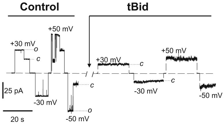
It was recently asserted that the voltage-dependent anion channel (VDAC) serves as a global regulator, or governor, of mitochondrial function (Lemasters and Holmuhamedov, Biochim Biophys Acta 1762:181-190, 2006). Indeed, VDAC, positioned on the interface between mitochondria and the cytosol (Colombini, Mol Cell Biochem 256:107-115, 2004), is at the control point of mitochondria life and death. This large channel plays the role of a "switch" that defines in which direction mitochondria will go: to normal respiration or to suppression of mitochondria metabolism that leads to apoptosis and cell death. As the most abundant protein in the mitochondrial outer membrane (MOM), VDAC is known to be responsible for ATP/ADP exchange and for the fluxes of other metabolites across MOM. It controls them by switching between the open and "closed" states that are virtually impermeable to ATP and ADP. This control has dual importance: in maintaining normal mitochondria respiration and in triggering apoptosis when cytochrome c and other apoptogenic factors are released from the intermembrane space into the cytosol. Emerging evidence indicates that VDAC closure promotes apoptotic signals without direct involvement of VDAC in the permeability transition pore or hypothetical Bax-containing cytochrome c permeable pores. VDAC gating has been studied extensively for the last 30 years on reconstituted VDAC channels. In this review we focus exclusively on physiologically relevant regulators of VDAC gating such as endogenous cytosolic proteins and mitochondrial lipids. Closure of VDAC induced by such dissimilar cytosolic proteins as pro-apoptotic tBid and dimeric tubulin is compared to show that the involved mechanisms are rather distinct. While tBid mostly modulates VDAC voltage gating, tubulin blocks the channel with the efficiency of blockage controlled by voltage. We also discuss how characteristic mitochondrial lipids, phospatidylethanolamine and cardiolipin, could regulate VDAC gating. Overall, we demonstrate that VDAC gating is not just an observation made under artificial conditions of channel reconstitution but is a major mechanism of MOM permeability control.


Figures

Fig. 1
Fig. 1
Schematics of VDAC gating. Upon VDAC closure a positively charged voltage sensor domain moves out of the channel to the membrane surface. This process is accompanied by decreasing of the pore volume and reversing of the channel selectivity. Closed states are impermeable for ATP/ADP fluxes but still conduct small ions and Ca2+. There are multiple stimuli which regulate VDAC gating or induce transient block of the open state
Fig. 2
Fig. 2
tBid-induced irreversible closure of VDAC. The current traces through the same single VDAC channel reconstituted into planar lipid membrane are shown before and after addition of 20 nM tBid in the cis-side of the membrane chamber. Membrane lipid composition: asolectin/DPhPC/CL/cholesterol; membrane bathing solution: 250 mM KCl, 1 mM CaCl2, 5 mM HEPES buffered at pH 7.2 (from Rostovtseva et al. 2004) (with permission ASBMB Journals)
Fig. 3
Fig. 3
VDAC gating asymmetry enhanced by non-lamellar mitochondrial lipids, PE and CL. a In the multichannel membranes formed from PE and PC + CL the closed state conductance is two times lower at negative potentials than at positive ones as follows from the conductance versus voltage plots normalized to the maximum conductance, Gmax (from Rostovtseva et al. 2006). Analysis of the plot also shows that for negative potentials the midpoint voltage, that is, the voltage at which half channels are open and half closed, is 10 mV less negative for PE as compared with PC. b A cartoon explaining gating asymmetry under lipid packing stress in PE bilayer. The model of VDAC gating was adopted from (Song et al. 1998a). There are one open and two sets of closed states of VDAC channel depending on the sign of the applied voltage. We hypothesize that the protein’s outer surface has a concave shape when channel adopts the closed state conformations at negative potentials. c Because of the concave shape, the lateral pressure in the hydrocarbon tail area of a planar bilayer shifts conformational equilibrium of VDAC channel in favor of the closed states of this shape. The probability to find these states in non-lamellar PE is much higher than in lamellar PC. Indeed, transition to the concave shape relieves the elastic stress of lipid packing, making the closed conformation energetically more favorable in membranes formed from non-lamellar lipid. A simple estimate illustrated here predicts a significant free energy change (see the text)
Fig. 4
Fig. 4
A model of tubulin-induced VDAC permeation block. One of tubulin negatively charged C-terminal tails partially blocks the channel conductance by entering VDAC pore in its open state and binding to the positively charged channel walls. This is seen on the traces of current through a single channel (bottom inset on the right) as tubulin-induced fast flickering of channel conductance between open and one closed state. This process is voltage-dependent and can be described by the first-order reaction. Interpolated to zero applied voltage the reaction is characterized by an equilibrium binding constant of the order of 1 nM −1. Analysis of the reaction voltage dependence gives an effective charge of about five elementary charges

References

    1. Andre N, Carre M, Brasseur G, Pourroy B, Kovacic H, Briand C, Braguer D. FEBS Lett. 2002;532:256–260. - PubMed
    1. Appaix F, Kuznetsov AV, Usson Y, Kay L, Andrienko T, Olivares J, Kaambre T, Sikk P, Margreiter R, Saks V. Exp Physiol. 2003;88:175–190. - PubMed
    1. Ardail D, Privat JP, Egretcharlier M, Levrat C, Lerme F, Louisot P. J Biol Chem. 1990;265:18797–18802. - PubMed
    1. Azoulay-Zohar H, Israelson A, Abu-Hamad S, Shoshan-Barmatz V. Biochem J. 2004;377:347–355. - PMC - PubMed
    1. Baines CP, Kaiser RA, Sheiko T, Craigen WJ, Molkentin JD. Nat Cell Biol. 2007;9:550–555. - PMC - PubMed

Publication types

MeSH terms