Review
. 2008:170:353-64.
doi: 10.1016/S0079-6123(08)00429-9.

Chronic stress plasticity in the hypothalamic paraventricular nucleus

Affiliations
Review

Chronic stress plasticity in the hypothalamic paraventricular nucleus

James P Herman et al. Prog Brain Res. 2008.

Abstract

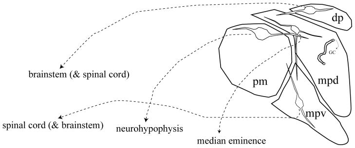
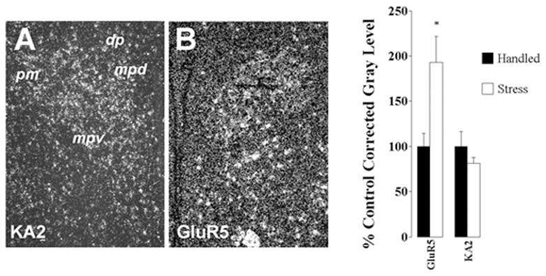
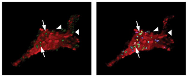
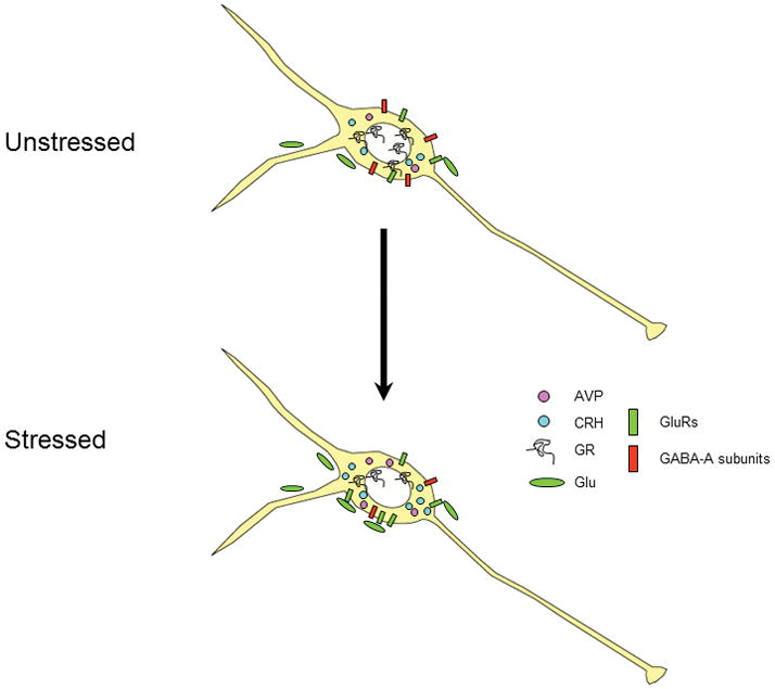
Proper integration and execution of the physiological stress response is essential for maintaining homoeostasis. Stress responses are controlled in large part by the paraventricular nucleus (PVN) of the hypothalamus, which contains three functionally distinct neural populations that modulate multiple stress effectors: (1) hypophysiotrophic PVN neurons that directly control the activity of the hypothalamic-pituitary-adrenocortical (HPA) axis; (2) magnocellular neurons and their secreted neurohypophysial peptides; and (3) brainstem and spinal cord projecting neurons that regulate autonomic function. Evidence for activation of PVN neurons during acute stress exposure demonstrates extensive involvement of all three effector systems. In addition, all PVN regions appear to participate in chronic stress responses. Within the hypophysiotrophic neurons, chronic stress leads to enhanced expression of secreted products, reduced expression of glucocorticoid receptor and GABA receptor subunits and enhanced glutamate receptor expression. In addition, there is evidence for chronic stress-induced morphological plasticity in these neurons, with chronic drive causing changes in cell size and altered GABAergic and glutamatergic innervation. The response of the magnocellular system varies with different chronic exposure paradigms, with changes in neurohypophysial peptide gene expression, peptide secretion and morphology seen primarily after intense stress exposure. The preautonomic cell groups are less well studied, but are likely to be associated with chronic stress-induced changes in cardiovascular function. Overall, the PVN is uniquely situated to coordinate responses of multiple stress effector systems in the face of prolonged stimulation, and likely plays a role in both adaptation and pathology associated with chronic stress.


Figures

Figure 1
Figure 1
Schematic of the organization of the hypothalamic paraventricular nucleus. Neurons in the dorsal parvocellular (dp) and ventral division of the medial parvocellular regions (mpv) project primarily to brainstem and spinal cord regions associated with autonomic control. Neurons present in the dorsal division of the medial parvocellular zone (mpd) project primarily to the median eminence and control ACTH release. The posterior magnocellular (pm) subdivision sends projections to the neurohypophysis. Note that the dendrites of neurons localized in all three regions ramify across subdivisions, allowing for intranuclear communication.
Figure 2
Figure 2
Distribution and regulation of KA2 and GluR5 receptor subunits in the PVN. As noted in A, KA2 mRNA is expressed in all subregions of the PVN (see Fig. 1 for abbreviations) GluR5 mRNA expression is particularly enriched in the PVNmpd. Semi-quantitative analysis of KA2 and GluR5 subunit mRNA expression in the PVN. Exposure to CVS causes a marked up-regulation of GluR5 expression in the PVN, whereas KA2 mRNA levels are unchanged.
Figure 3
Figure 3
3-D Rendering of Confocal Images: CRH and VGLUT2. Left: cropped, 3-D rendering of a single CRH cell (red) amid green VGlut2 puncta, indicative of glutamatergic terminals. Right: processed image of the same cell, with multi-colored boutons indicating those showing significant overlap between red and green fluorescence signal. Arrows indicate examples of boutons that ‘contact’ the CRH neuron, as determined by the ‘overlap’ algorithm in Volocity 2; boutons that do not contact (arrowheads) do not register as overlapping entities. The volumes of positive elements in the cell and bouton channels were measured independently, using the classification features; overlap was determined using the co-localization feature, which determines incidence and volume of boutons contacting the immunolabeled cell. Preliminary analysis of ADX data using 6-cells/group (2 each from 3 animals/group) revealed a 56% increase in VGlut2 bouton counts/cell surface area using this method. In addition, ADX animals showed an increase in contact bouton overlap area and in overall bouton size.
Figure 4
Figure 4
Schematic diagram of multiple neuroplastic changes in the PVNmpd under conditions of chronic drive. Following chronic stress, there is increased expression of ACTH secretagogs CRH and AVP. At the same time, expression of the glucocorticoid receptor (GR) is down-regulated, suggesting loss of feedback capacity. Expression of the GABA-A receptor subunits beta-1 and beta-2 are decreased, consistent with reduced GABAergic inhibition. In contrast, expression of the kainate preferring GluR5 subunit mRNA is increased, suggesting increased glutamate signaling via non-NMDA receptors. Finally, glutamatergic innervation of CRH neurons is markedly increased following chronic drive by adrenalectomy, suggesting the potential for stress-induced synaptic plasticity.

References

    1. Albeck DS, McKittrick CR, Blanchard DC, Blanchard RJ, Nikulina J, McEwen BS, Sakai RR. Chronic social stress alters levels of corticotropin-releasing factor and arginine vasopressin mRNA in rat brain. J Neurosci. 1997;17:4895–903. - PMC - PubMed
    1. Antoni FA. Hypothalamic control of adrenocorticotropin secretion: Advances since the discovery of 41-residue corticotropin-releasing factor. Endocrine Rev. 1986;7:351–378. - PubMed
    1. Carrasco M, Portillo F, Larsen PJ, Vallo JJ. Insulin and glucose administration stimulates Fos expression in neurones of the paraventricular nucleus that project to autonomic preganglionic structures. J Neuroendocrinol. 2001;13:339–46. - PubMed
    1. Chappell PB, Smith MA, Kilts CD, Bissette G, Ritchie J, Anderson C, Nemeroff CB. Alterations in corticotropin-releasing factor-like immunoreactivity in discrete brain regions after acute and chronic stress. J Neurosci. 1986;6:2908–2914. - PMC - PubMed
    1. Cullinan WE. GABA(A) receptor subunit expression within hypophysiotropic CRH neurons: a dual hybridization histochemical study. J Comp Neurol. 2000;419:344–51. - PubMed

Publication types

MeSH terms