. 2008:170:589-601.
doi: 10.1016/S0079-6123(08)00446-9.

Potential utility of aquaporin modulators for therapy of brain disorders

Affiliations

Potential utility of aquaporin modulators for therapy of brain disorders

Marios C Papadopoulos et al. Prog Brain Res. 2008.

Abstract

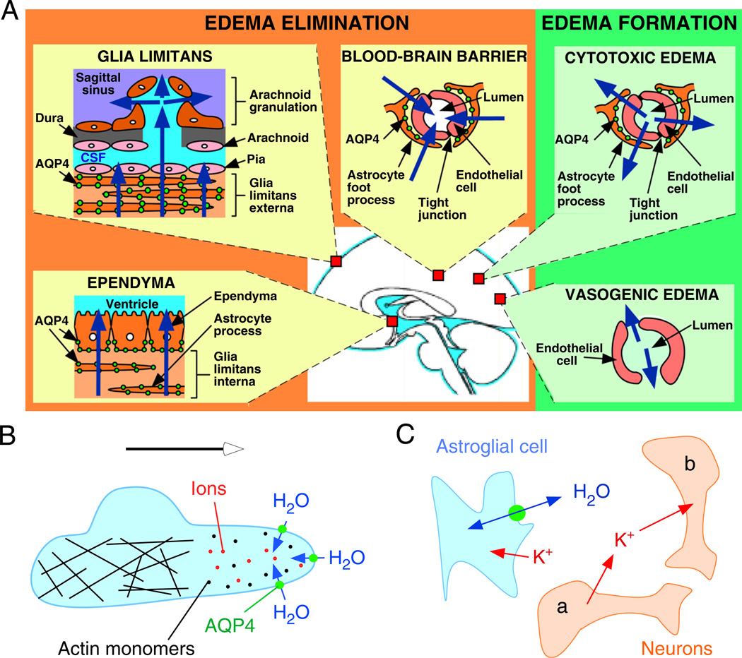
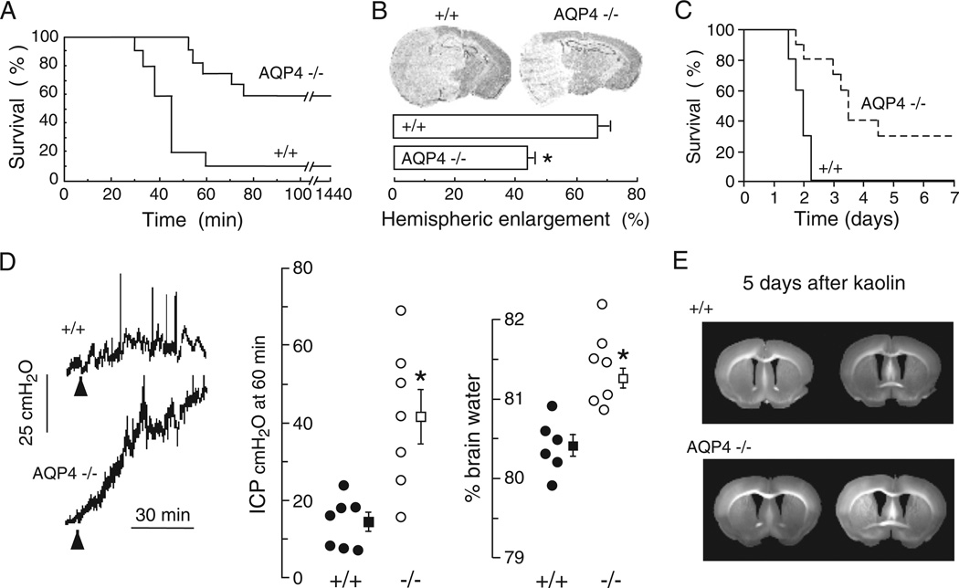
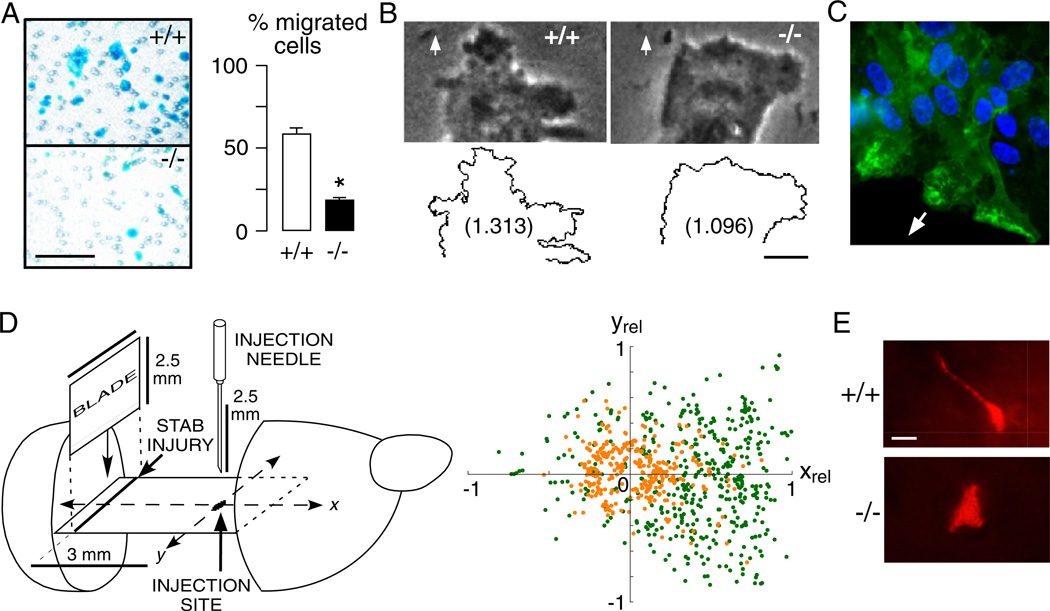
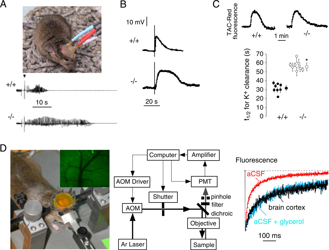
Of the several aquaporin (AQP) water channels expressed in the central nervous system, AQP4 is an attractive target for drug discovery. AQP4 is expressed in astroglia, most strongly at the blood-brain and brain-cerebrospinal fluid barriers. Phenotype analysis of AQP4 knockout mice indicates the involvement of AQP4 in three distinct processes: brain water balance, astroglial cell migration and neural signal transduction. By slowing water uptake into the brain, AQP4 knockout mice manifest reduced brain swelling and improved outcome in models of cytotoxic cerebral oedema such as water intoxication, focal ischaemia and meningitis. However, by slowing the clearance of excess water from brain, AQP4 knockout mice do worse in models of vasogenic oedema such as brain tumour, abscess and hydrocephalus. AQP4 deficient astroglial cells show greatly impaired migration in response to chemotactic stimuli, reducing glial scar formation, by a mechanism that we propose involves AQP4-facilitated water flux in lamellipodia of migrating cells. AQP4 knockout mice also manifest increased seizure threshold and duration, by a mechanism that may involve slowed K(+) uptake from the extracellular space (ECS) following neuroexcitation, as well as ECS expansion. Notwithstanding challenges in drug delivery to the central nervous system and their multiplicity of actions, AQP4 inhibitors have potential utility in reducing cytotoxic brain swelling, increasing seizure threshold and reducing glial scar formation; enhancers of AQP4 expression have potential utility in reducing vasogenic brain swelling. AQP4 modulators may thus offer new therapeutic options for stroke, tumour, infection, hydrocephalus, epilepsy and traumatic brain and spinal cord injury.


Figures

Fig. 1
Fig. 1
Schematic depicting three distinct roles of AQP4 (green circles) in brain function: (A) brain water balance, (B) astroglial cell migration and (C) neuronal excitation. (A) Green. Routes of oedema formation in the two types of brain oedema (cytotoxic — through AQP4, vasogenic — through interendothelial spaces). Orange. Oedema fluid is eliminated by AQP4 through the glial limitans into subarachnoid CSF, through ependyma and sub-ependymal astroglia into ventricular CSF, and through astroglial pericapillary foot processes into blood. (B) AQP4 polarizes to the leading edge of migrating astroglia and accelerates cell migration. AQP4 facilitates water entry into lamellipodial protrusions in response to intracellular hyperosmolality produced by actin depolymerization and ion influx. (C) AQP4 deletion reduces neuroexcitation. Active neurons (neuron a) release K+ into the extracellular space (ECS). Increased extracellular [K+] depolarizes quiescent neurons (neuron b). AQP4 deletion increases ECS volume and reduces astroglial cell K+ reuptake. This buffers the increase in extracellular [K+] by active neuron a, preventing depolarization of quiescent neuron b. See text for further explanations. (See Color Plate 46.1 in color plate section.)
Fig. 2
Fig. 2
AQP4 deletion reduces brain water accumulation in cytotoxic oedema, but slows removal of excess brain water in vasogenic oedema. (A) Water intoxication model of cytotoxic oedema. Survival of 12 wildtype vs. 12 AQP4 knockout mice after acute water intoxication produced by intraperitoneal water injection (20% body weight). (B) (Top) Ischaemic stroke model of cytotoxic oedema. Brain sections of mice at 24 h after ischaemic stroke produced by permanent middle cerebral artery occlusion. (Bottom) Average hemispheric enlargement expressed as a percentage determined by image analysis of brain sections (7 AQP4+/+ vs. 7 AQP4−/− mice, SEM, * P < 0.0002). (C) Mouse survival (10 AQP4+/+ vs. 10 AQP4−/− mice, P < 0.001) in a bacterial model of meningitis produced by cisternal injection of S. pneumoniae. (D) Reduced elevation in intracranial pressure (ICP, SEM, * P < 0.01) and brain water content (SEM, * P < 0.001) following continuous intraparenchymal infusion of artificial cerebrospinal fluid at 0.5 µL/min. (E) Accelerated progression of hydrocephalus in AQP4 null mice. Coronal sections wildtype and AQP4 null mouse brain at 5 days after kaolin injection. Data from Manley et al., 2000, Papadopoulos et al., 2004 and Bloch et al., 2006.
Fig. 3
Fig. 3
AQP4 facilitates astroglial migration in vitro and in brain. (A) Left. Boyden chamber migration assay showing AQP4+/+ and AQP4−/− astroglia (blue) after scraping off the non-migrated cells. Astroglia were plated on the top chamber of porous transwell filter (2.8 × 10⁴/cm2) and were allowed to migrate for 6 h towards 10% FBS as chemoattractant. Bar, 100 µm. Right. Summary of migration experiments (SEM, * P < 0.001). (B) Phase contrast micrographs (Top) and outline (Bottom) of the leading end of a migrating AQP4+/+ and AQP4−/− astroglial cell in the in vitro wound assay. Arrows show direction of migration. Numbers are fractal dimensions (larger number denotes more irregular cell membrane). Bar 10 µm. (C) AQP4 protein (green) polarization to the front end of migrating astroglia in a wound assay. Arrow shows direction of migration. (D) Left. Stab injury/cell injection model of astroglial migration in mouse brain. Two days before cell injection, a stab was created as shown. Cultured AQP4+/+ and AQP4+/+ astroglia were fluorescently labelled and injected as indicated. Right. Locations of migrating fluorescently stained AQP4+/+ (green) and AQP4−/− (orange) astroglia. The x-axis is relative distance between injection and stab injury sites (xrel). (E) High magnification fluorescence micrographs of migrating AQP4+/+ and AQP4−/− astroglia. Bar 5 µm. Data from Saadoun et al., 2005b and Auguste et al., 2007. (See Color Plate 46.3 in color plate section.)
Fig. 4
Fig. 4
AQP4 involvement in brain neuroexcitation. (A) Increased seizure duration in AQP4 null mice. Top. Bipolar electrodes implanted in the right hippocampus were connected to a stimulator and electroencephalograph recording system. Bottom. Representative electroencephalograms in freely moving mice following electrically induced generalized seizures. (B) Delayed K+ clearance in brain following electrically induced seizure-like neuroexcitation. Measurements done using K+-sensitive microelectrodes inserted into brain cortex in living mice. (C) Slowed K+ clearance in brain ECS during cortical spreading depression measured using TAC-Red, a K+-sensitive fluorescent probe. Top. Representative data. Bottom. Half-times (t1/2) for K+ reuptake. (D) Expanded brain ECS in AQP4 null mice measured by cortical surface photobleaching. Left. Mouse brain surface exposed to FITC-dextran with dura intact following craniectomy and fluorescence imaging of cortical surface after loading with FITC-dextran (inset). Middle. Photobleaching apparatus. A laser beam is modulated by an acousto-optic modulator and directed onto the surface of the cortex using a dichroic mirror and objective lens. Right. In vivo fluorescence recovery in cortex of wildtype mouse shown in comparison to aCSF and 30% glycerol in aCSF. Taken from Binder et al., 2006, Binder et al., 2004b and Padmawar et al., 2005. (See Color Plate 46.4 in color plate section.)

References

    1. Amiry-Moghaddam M, Lindland H, Zelenin S, Roberg BA, Gundersen BB, Petersen P, Rinvik E, Torgner IA, Ottersen OP. Brain mitochondria contain aquaporin water channels: evidence for the expression of a short AQP9 isoform in the inner mitochondrial membrane. Faseb J. 2005;19:1459–1467. - PubMed
    1. Amiry-Moghaddam M, Otsuka T, Hurn PD, Traystman RJ, Haug FM, Froehner SC, Adams ME, Neely JD, Agre P, Ottersen OP, Bhardwaj A. An alpha-syntrophin-dependent pool of AQP4 in astroglial end-feet confers bidirectional water flow between blood and brain. Proc. Natl. Acad. Sci. U.S.A. 2003a;100:2106–2111. - PMC - PubMed
    1. Amiry-Moghaddam M, Williamson A, Palomba M, Eid T, de Lanerolle NC, Nagelhus EA, Adams ME, Froehner SC, Agre P, Ottersen OP. Delayed K+ clearance associated with aquaporin-4 mislocalization: phenotypic defects in brains of alpha-syntrophin-null mice. Proc. Natl. Acad. Sci. U.S.A. 2003b;100:13615–13620. - PMC - PubMed
    1. Amiry-Moghaddam M, Xue R, Haug FM, Neely JD, Bhardwaj A, Agre P, Adams ME, Froehner SC, Mori S, Ottersen OP. Alpha-syntrophin deletion removes the perivascular but not endothelial pool of aquaporin-4 at the blood–brain barrier and delays the development of brain edema in an experimental model of acute hyponatremia. Faseb J. 2004;18:542–544. - PubMed
    1. Aoki K, Uchihara T, Tsuchiya K, Nakamura A, Ikeda K, Wakayama Y. Enhanced expression of aquaporin 4 in human brain with infarction. Acta Neuropathol. (Berl.) 2003;106:121–124. - PubMed

MeSH terms