. 2008 Aug;83(2):208-18.
doi: 10.1016/j.ajhg.2008.07.007. Epub 2008 Jul 24.

Evolutionary forces shape the human RFPL1,2,3 genes toward a role in neocortex development

Affiliations

Evolutionary forces shape the human RFPL1,2,3 genes toward a role in neocortex development

Jérôme Bonnefont et al. Am J Hum Genet. 2008 Aug.

Abstract

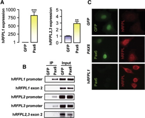
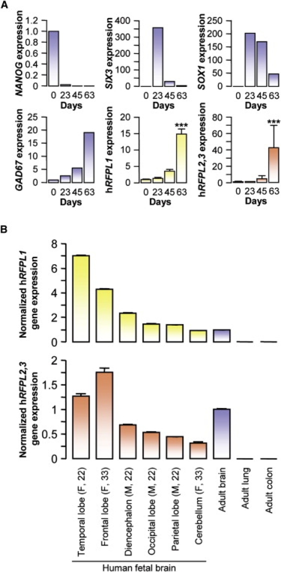
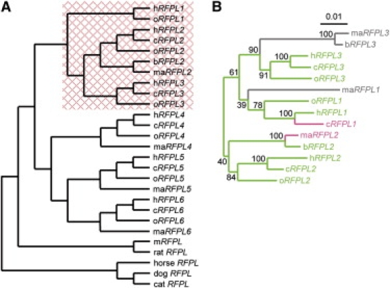
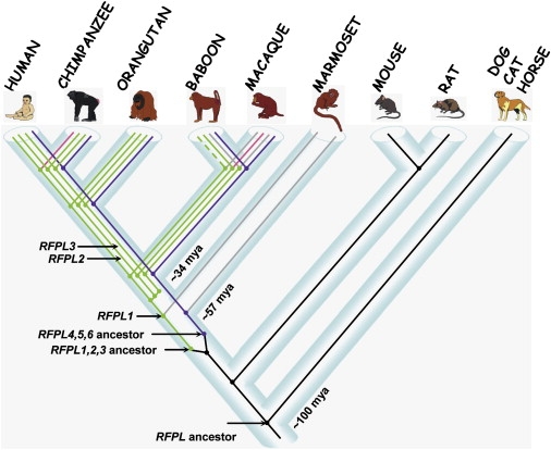
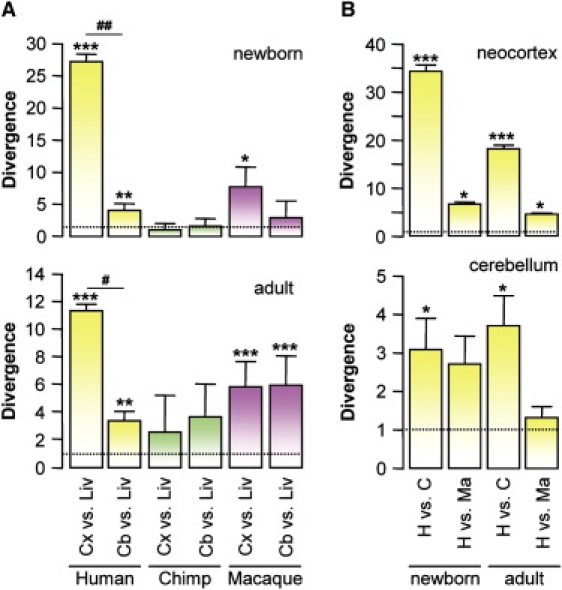
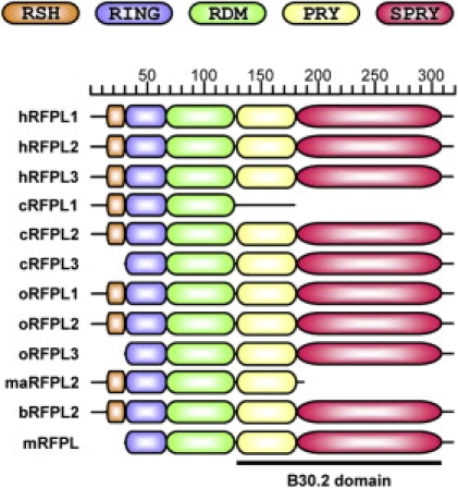
The size and organization of the brain neocortex has dramatically changed during primate evolution. This is probably due to the emergence of novel genes after duplication events, evolutionary changes in gene expression, and/or acceleration in protein evolution. Here, we describe a human Ret finger protein-like (hRFPL)1,2,3 gene cluster on chromosome 22, which is transactivated by the corticogenic transcription factor Pax6. High hRFPL1,2,3 transcript levels were detected at the onset of neurogenesis in differentiating human embryonic stem cells and in the developing human neocortex, whereas the unique murine RFPL gene is expressed in liver but not in neural tissue. Study of the evolutionary history of the RFPL gene family revealed that the RFPL1,2,3 gene ancestor emerged after the Euarchonta-Glires split. Subsequent duplication events led to the presence of multiple RFPL1,2,3 genes in Catarrhini ( approximately 34 mya) resulting in an increase in gene copy number in the hominoid lineage. In Catarrhini, RFPL1,2,3 expression profile diverged toward the neocortex and cerebellum over the liver. Importantly, humans showed a striking increase in cortical RFPL1,2,3 expression in comparison to their cerebellum, and to chimpanzee and macaque neocortex. Acceleration in RFPL-protein evolution was also observed with signs of positive selection in the RFPL1,2,3 cluster and two neofunctionalization events (acquisition of a specific RFPL-Defining Motif in all RFPLs and of a N-terminal 29 amino-acid sequence in catarrhinian RFPL1,2,3). Thus, we propose that the recent emergence and multiplication of the RFPL1,2,3 genes contribute to changes in primate neocortex size and/or organization.


Figures

Figure 1
Figure 1
Pax6 Interacts with the hRFPL1,2,3 Gene Promoters and Induces Their Transcript and Protein Expressions (A) hRFPL1 and hRFPL2,3 expressions were determined by real-time PCR after overexpression of GFP or Pax6 in HeLa cells. Transcript levels are indicated as the fold increase relative to the control level (GFP-transduced cells). hRFPL2 and hRFPL3 are detected by a common set of primers. Results are displayed as mean ± standard error (SEM). Statistical analyses were done with Student's t test, ∗∗p < 0.01 and ∗∗∗p < 0.001. (B) In vivo binding of Pax6 to hRFPL1,2,3 promoters was assessed by chromatin-immunoprecipitation assay. After chromatin immunoprecipitation with a Pax6 antibody, endpoint PCRs were performed with primers specific for each hRFPL1,2,3 promoter or for exon 2 of each gene for visualization of nonspecific immunoprecipitation. “Input” represents dilutions of input chromatin used as PCR controls. (C) Immunocytochemical detection of hRFPL1,2,3 proteins in GFP- and Pax6-expressing HeLa cells with a pan-hRFPL1,2,3 antibody. hRFPL1 overexpression was used as a control for antibody specificity.
Figure 2
Figure 2
hRFPL1,2,3 Genes Are Expressed during Human Neurogenesis In Vitro and In Vivo (A) hRFPL1 and hRFPL2,3 expressions were determined by real-time PCR during human embryonic-stem-cells-derived neurogenesis and normalized to the cDNA level in fetal brain. Results are displayed as mean ± standard error (SEM). ∗∗∗p < 0.001 versus day 0 with one-way ANOVA followed by post hoc Tukey's test. hRFPL1 and hRFPL2,3 expressions were correlated to those of the neuronal marker GAD67 (R2 = 0.99, p = 0.003 for hRFPL1 versus GAD67; R2 = 0.98, p = 0.012 for hRFPL2,3 versus GAD67 with Pearson product moment correlation). (B) hRFPL1 and hRFPL2,3 expressions were assessed in different structures of the developing brain and in adult neocortex by real-time PCR normalized to the level of expression observed in adult brain. The gender of the embryo (M, male; F, female) and its age in weeks are indicated in the parentheses. Results are displayed as mean ± standard error (SEM).
Figure 3
Figure 3
Topology of the RFPL Gene Family (A) Phylogenetic tree based on RFPL coding sequences encoding the likely functional proteins in Laurasiatheria, Glires, and Catarrhini. The RFPL1,2,3 clade shows a significant (p < 0.0001) positive selection or constraint relaxation that is indicated by the red box. (B) ML phylogenetic tree of the catarrhinian RFPL1,2,3 genes. The topology and branch lengths are based on concatenated alignments of the intron and the second exon. Branch lengths are scaled to the number of substitutions per site. Bootstrap support values are indicated at each node. Likely functional genes are shown in green, truncated genes are shown in pink, and nonfunctional genes are shown in gray. The following abbreviations are used in both panels: h, human; c, chimpanzee; o, orangutan; b, baboon; ma, macaque; and m, mouse.
Figure 4
Figure 4
Timing of the Emergence of the RFPL Genes during Evolution The RFPL gene ancestor is shown in black. Each likely functional RFPL1, RFPL2, and RFPL3 gene is indicated in green, truncated genes are indicated in pink, and genes with no ORF are indicated in gray. Putative baboon RFPL1 gene is indicated in a dashed line. The RFPL4,5,6 gene cluster is represented by a single purple line. The estimated time of the gene-ancestor emergence and subsequent duplication events are indicated in millions of years (mya).
Figure 5
Figure 5
Expression Divergence between Human, Chimpanzee and Macaque RFPL1,2,3 Genes in Brain RFPL1,2,3 gene expressions were assessed in newborn and adult neocortex, cerebellum, and liver by microarray transcriptome studies in humans, chimpanzees, and macaques. Results are displayed as mean ± standard error (SEM). The following abbreviations are used: Cx, neocortex; Cb, cerebellum; Liv, liver; H, human; C, chimpanzee; and Ma, macaque. (A) shows intraspecies divergence in RFPL1,2,3 tissue expression. Cortical and cerebellar RFPL1,2,3 transcript levels were normalized to that of the liver. p < 0.05, ∗∗p < 0.01, and ∗∗∗p < 0.001 for neocortex or cerebellum versus liver; #p < 0.05 and ##p < 0.01 for neocortex versus cerebellum with one-way ANOVA followed by post hoc Tukey test. (B) shows interspecies divergence in cortical and cerebellar RFPL1,2,3 expression levels. hRFPL1,2,3 expressions were normalized to those in chimpanzee or macaque. p < 0.05 and ∗∗∗p < 0.001 with Student's t test.
Figure 6
Figure 6
RFPL-Protein-Domain Predictions Reveal the Acquisition of RDM and RSH as Neofunctionalization Events Identification of RFPL-specific RDM (RFPL-defining motif) and RSH (RFPL1,2,3-specifying helix) were obtained after the alignment of the likely functional catarrhinian RFPL1,2,3 and murine mRFPL proteins and in silico predictions of secondary structures and protein domains. The following abbreviations are used: h, human; c, chimpanzee; o, orangutan; b, baboon; ma, macaque; and m: mouse.

References

    1. Kaas J.H. From mice to men: The evolution of the large, complex human brain. J. Biosci. 2005;30:155–165. - PubMed
    1. Kaas J.H. Evolution of the neocortex. Curr. Biol. 2006;16:R910–R914. - PubMed
    1. Gotz M., Huttner W.B. The cell biology of neurogenesis. Nat. Rev. Mol. Cell Biol. 2005;6:777–788. - PubMed
    1. Kornack D.R., Rakic P. Changes in cell-cycle kinetics during the development and evolution of primate neocortex. Proc. Natl. Acad. Sci. USA. 1998;95:1242–1246. - PMC - PubMed
    1. Kornack D.R. Neurogenesis and the evolution of cortical diversity: Mode, tempo, and partitioning during development and persistence in adulthood. Brain Behav. Evol. 2000;55:336–344. - PubMed

Publication types

LinkOut - more resources