. 2008 Sep 1;378(2):243-53.
doi: 10.1016/j.virol.2008.05.007. Epub 2008 Jul 25.

The linker domain of poly(rC) binding protein 2 is a major determinant in poliovirus cap-independent translation

Affiliations

The linker domain of poly(rC) binding protein 2 is a major determinant in poliovirus cap-independent translation

Polen Sean et al. Virology. .

Abstract

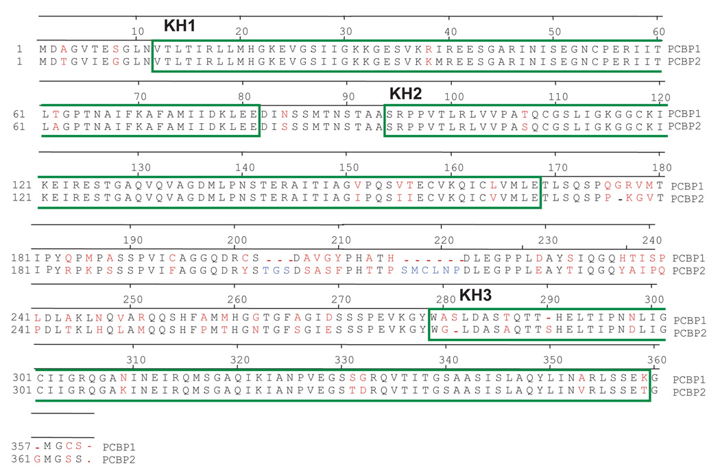
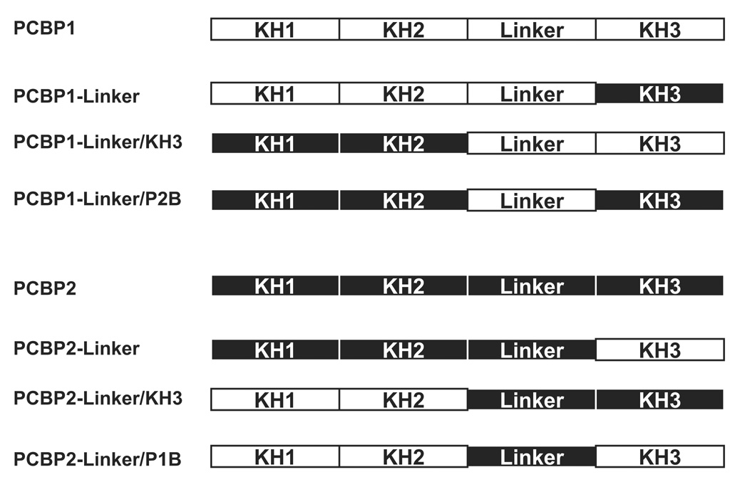
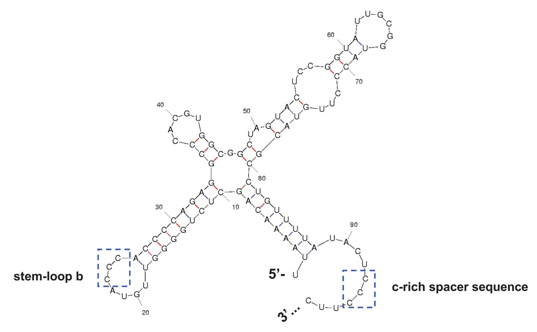
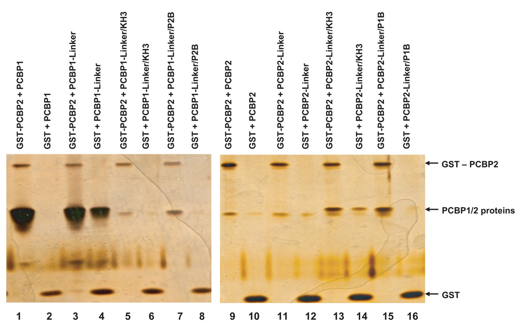
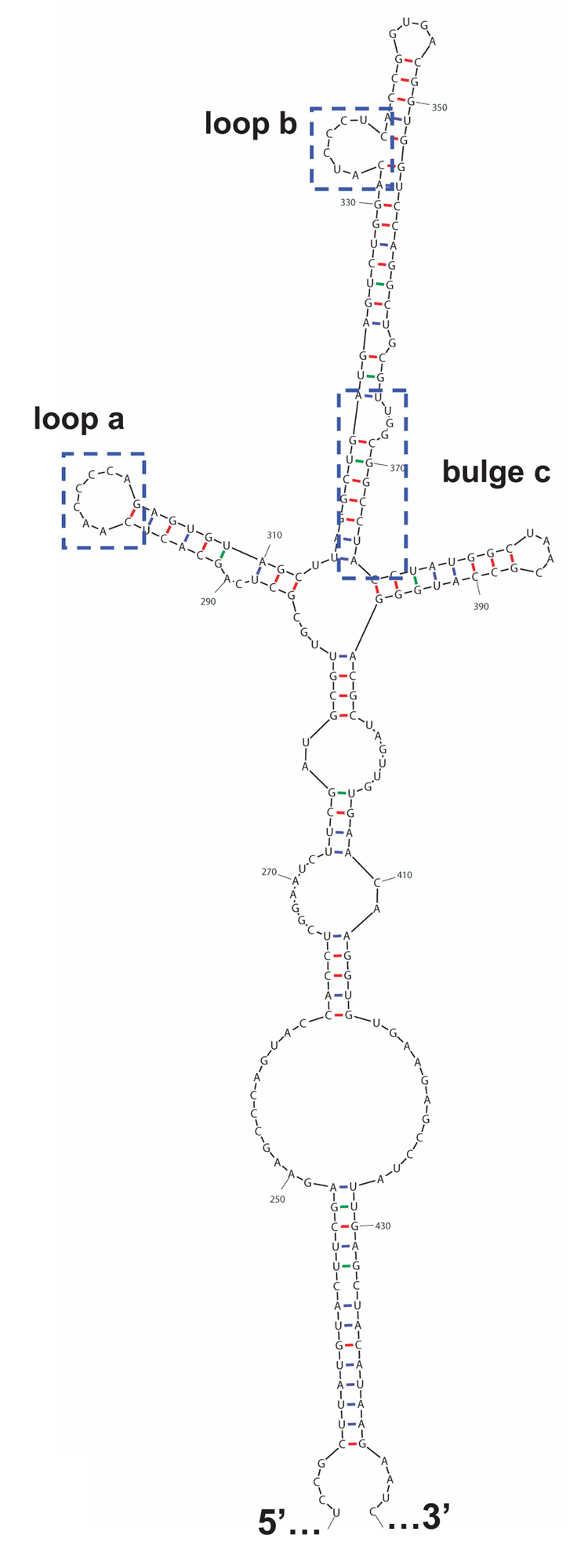
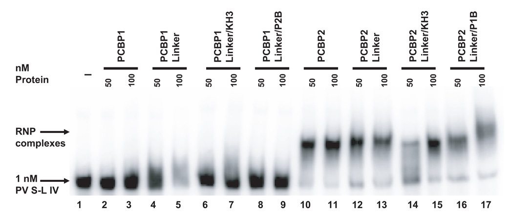
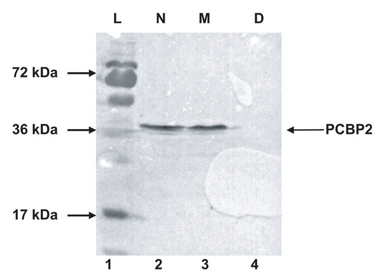
Poliovirus, a member of the enterovirus genus in the family Picornaviridae, is the causative agent of poliomyelitis. Translation of the viral genome is mediated through an internal ribosomal entry site (IRES) encoded within the 5' noncoding region (5' NCR). IRES elements are highly structured RNA sequences that facilitate the recruitment of ribosomes for translation. Previous studies have shown that binding of a cellular protein, poly(rC) binding protein 2 (PCBP2), to a major stem-loop structure in the genomic 5' NCR is necessary for the translation of picornaviruses containing type I IRES elements, including poliovirus, coxsackievirus, and human rhinovirus. PCBP1, an isoform that shares approximately 90% amino acid identity to PCBP2, cannot efficiently stimulate poliovirus IRES-mediated translation, most likely due to its reduced binding affinity to stem-loop IV within the poliovirus IRES. The primary differences between PCBP1 and PCBP2 are found in the so-called linker domain between the second and third K-homology (KH) domains of these proteins. We hypothesize that the linker region of PCBP2 augments binding to poliovirus stem-loop IV RNA. To test this hypothesis, we generated six PCBP1/PCBP2 chimeric proteins. The recombinant PCBP1/PCBP2 chimeric proteins were able to interact with poliovirus stem-loop I RNA and participate in protein-protein interactions. We demonstrated that the PCBP1/PCBP2 chimeric proteins with the PCBP2 linker, but not with the PCBP1 linker, were able to interact with poliovirus stem-loop IV RNA, and could subsequently stimulate poliovirus IRES-mediated translation. In addition, using a monoclonal anti-PCBP2 antibody (directed against the PCBP2 linker domain) in mobility shift assays, we showed that the PCBP2 linker domain modulates binding to poliovirus stem-loop IV RNA via a mechanism that is not inhibited by the antibody.


Figures

Figure 1
Figure 1. Domain compositions of PCBP1/PCBP2 chimeric proteins
A. Amino acid sequence alignment of PCBP1 and PCBP2. The amino acid sequences of the KH domains are boxed in green. The amino acid sequences of KH1 and KH2 are nearly identical, with slight differences in KH3 and major differences in the linker region between the KH2 and KH3 domain. Amino acid differences are denoted in red and amino acids that only exist in the PCBP2 linker are denoted in blue. Dashes indicate the absence of a corresponding amino acid in one protein or the other. B. Schematic of the PCBP1/PCBP2 chimeric proteins. The proteins were constructed using restriction sites that were introduced at the interface of the KH2/linker (XhoI) and linker/KH3 (NheI) into a pET22 expression plasmid. KH domain and linker sequences from PCBP1 are indicated by white boxes while those from PCBP2 are indicated by black boxes.
Figure 2
Figure 2. Characterization of PCBP1/PCBP2 chimeric proteins
A. Mfold predicted RNA secondary structure of stem-loop I. The nucleotide constraints were determined by chemical and enzymatic structure probing (Skinner et al., 1989). The structure shown represents nucleotide 1 to 98 for the poliovirus type 1 genomic RNA. Poliovirus stem-loop I is predicted to form a cloverleaf-like structure. Stem-loop b and the c-rich spacer sequence, boxed, have been shown to interact with PCBP1/2 (Parsley et al., 1997; Toyoda et al., 2007). B. Electrophoretic mobility shift assay of PCBP1/PCBP2 chimeric proteins with poliovirus stem-loop I RNA. In vitro transcribed [32P] UTP-labeled poliovirus stem-loop I RNA at a final concentration of 0.1 nM was incubated with 50 or 100 nM of purified recombinant PCBP1/PCBP2 chimeric protein for 10 minutes at 30°C, and the reaction was resolved by native polyacrylamide gel electrophoresis. Lanes 1 and 16 show the RNA in the absence of protein. Lanes 2–5, 8–15 and 17–18 show ribonucleoprotein (RNP) complexes formed by the interaction of the PCBP1/PCBP2 chimeric proteins with the RNA. The RNP complexes are denoted by the upper arrow. The samples in lanes 16–18 were subjected to electrophoresis for an additional hour (compared to those samples in lanes 1–15), thus accounting for the increased distance between the RNP complex and the free RNA. Lanes 6–7 show that the PCBP1-Linker/P2B chimera is unable to interact with poliovirus stem-loop I RNA.
Figure 3
Figure 3. GST pull-down assays of GST-PCBP2 and PCBP1/PCBP2 chimeric proteins
GST-PCBP2 (2 µg) and His-tagged PCBP1/PCBP2 chimeric proteins (5 µg) were subjected to pull-down assays as described in Materials and Methods. Lanes 1–16 show the variable interaction of GST-PCBP2 with the PCBP1/PCBP2 chimeric proteins over GST alone (i.e., non-specific background interactions). Lanes 1–2 and 9–10 show the interactions of wild-type PCBP1 and PCBP2 with GST-PCBP2 and GST (control), respectively.
Figure 4
Figure 4. The PCBP2 linker mediates binding to poliovirus stem-loop IV RNA
A. Mfold predicted RNA secondary structure of stem-loop IV. Nucleotide constraints were determined by chemical and enzymatic structure probing (Skinner et al., 1989). The structure shown represents nucleotide 230 to 444 for poliovirus type 1 genomic RNA. Poliovirus stem-loop IV is predicted to form a cruciform-like structure. Loops a and b, and bulge c, boxed, have been identified to interact with PCBP2 (Gamarnik and Andino, 2000). B. Electrophoretic mobility shift assay of PCBP1/PCBP2 chimeric proteins with poliovirus stem-loop IV RNA. In vitro transcribed [32P] UTP-labeled poliovirus stem-loop IV RNA at a final concentration of 1 nM was incubated with 50 or 100 nM of purified recombinant PCBP1/PCBP2 chimeric proteins for 10 minutes at 30°C, and the reaction was resolved by native polyacrylamide gel electrophoresis. Lane 1 shows the electrophoretic mobility of the RNA probe in the absence of added protein. Lanes 2–9 show the incubation of the PCBP1-linker containing chimeric proteins with the RNA. Lanes 10–17 show the formation of ribonucleoprotein complexes by the interaction of the PCBP2-linker containing chimeric proteins with the RNA. The RNP complexes are denoted by the upper arrow.
Figure 5
Figure 5. Addition of PCBP2-linker containing chimeric proteins to PCBP-depleted cytoplasmic extracts rescues poliovirus IRES-mediated translation
A. Western blot analysis of HeLa S10 cytoplasmic extract depleted of PCBP. A total of 50 µg of protein from HeLa cell cytoplasmic extract or HeLa cell cytoplasmic extract depleted of PCBPs via poly(rC)-sepharose affinity chromatography was resolved by SDS-PAGE, transferred to a PVDF membrane, and probed using a monoclonal anti-PCBP2 antibody. Lane 1 shows the molecular weight ladder, L. Lanes 2–4 display the non-depleted HeLa S10 (N), mock-depleted HeLa S10 (M) and PCBP-depleted HeLa S10 (D). B. Luciferase assays measuring the in vitro translation of PV 5′ NCR-luc RNA with PCBP1/PCBP2 chimeric proteins. We incubated 50 fmol of PV 5′ NCR-luc RNA and 100 nM of PCBP1/PCBP2 chimeric proteins in PCBP-depleted HeLa S10 cytoplasmic extract for 2.5 hours at 30°C and assayed for luciferase activity. The relative light unit (RLU) values are averages from three separate reactions. The error bars are standard deviations from the mean.
Figure 6
Figure 6. Monoclonal antibody targeting the linker region of PCBP2 does not interfere with binding to stem-loop IV RNA
A. Purified, recombinant PCBP1/PCBP2 chimeric proteins (1 µg) were resolved by SDS-PAGE and stained with coomassie blue (upper panel). Western blot analysis of the PCBP1/PCBP2 chimeric proteins (lower panel). PCBP1/PCBP2 chimeric proteins (1µg) were resolved by SDS-PAGE, transferred to a PVDF membrane, and probed with a monoclonal anti-PCBP2 antibody. Only proteins that contain the PCBP2 linker are detected by the antibody (shown by the arrow). B. Mobility shift assay of recombinant PCBP2 and poliovirus stem-loop IV RNA with the addition of a monoclonal or polyclonal anti-PCBP2 IgG. In vitro transcribed [32P] UTP-labeled poliovirus stem-loop IV RNA at a final concentration of 1 nM was incubated with 50 or 100 nM of purified recombinant PCBP2 for 10 minutes at 30°C. 1 µg of antibody was then added to the reaction and incubated for an additional 10 minutes at 30°C. The reaction was then resolved by native polyacrylamide gel electrophoresis. Lane 1 is the RNA alone. Lanes 2–3 show the incubation of the RNA in the presence of the monoclonal or polyclonal anti-PCBP2 IgG. Lanes 4–5 show ribonucleoprotein complexes formed by the interaction of PCBP2 with the RNA, as indicated by the PCBP2/ stem-loop IV RNP complexes arrow. Lanes 6–7 show the super-shifted RNP complex mediated by the monoclonal anti-PCBP2 antibody, as indicated by the super-shifted RNP complex arrow. Lanes 8–9 show that the polyclonal anti-PCBP2 antibody did not super-shift the PCBP2-poliovirus stem-loop IV RNP complex.

Similar articles

Cited by

References

    1. Andino R, Rieckhof GE, Baltimore D. A functional ribonucleoprotein complex forms around the 5' end of poliovirus RNA. Cell. 1990;63:369–380. - PubMed
    1. Barton DJ, Flanegan JB. Coupled translation and replication of poliovirus RNA in vitro: synthesis of functional 3D polymerase and infectious virus. J Virol. 1993;67:822–831. - PMC - PubMed
    1. Bedard KM, Daijogo S, Semler BL. A nucleo-cytoplasmic SR protein functions in viral IRES-mediated translation initiation. EMBO J. 2007;26:459–467. - PMC - PubMed
    1. Bedard KM, Walter BL, Semler BL. Multimerization of poly(rC) binding protein 2 is required for translation initiation mediated by a viral IRES. RNA. 2004;10:1266–1276. - PMC - PubMed
    1. Belsham GJ, Sonenberg N. RNA-protein interactions in regulation of picornavirus RNA translation. Microbiol. Rev. 1996;60:499–511. - PMC - PubMed

Publication types

MeSH terms