

Macrophage roles following myocardial infarction
- PMID: 18656272
- PMCID: PMC2857604
- DOI: 10.1016/j.ijcard.2008.04.059
Macrophage roles following myocardial infarction
Abstract
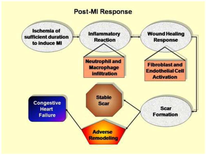
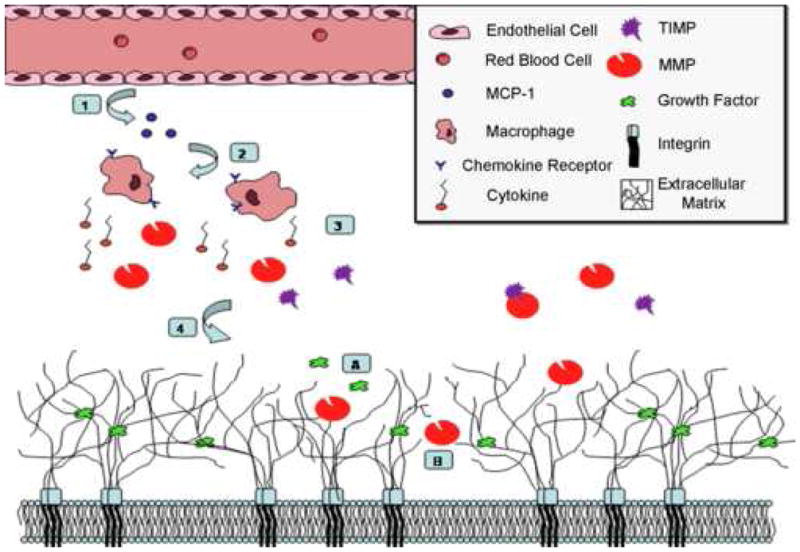
Following myocardial infarction (MI), circulating blood monocytes respond to chemotactic factors, migrate into the infarcted myocardium, and differentiate into macrophages. At the injury site, macrophages remove necrotic cardiac myocytes and apoptotic neutrophils; secrete cytokines, chemokines, and growth factors; and modulate phases of the angiogenic response. As such, the macrophage is a primary responder cell type that is involved in the regulation of post-MI wound healing at multiple levels. This review summarizes what is currently known about macrophage functions post-MI and borrows literature from other injury and inflammatory models to speculate on additional roles. Basic science and clinical avenues that remain to be explored are also discussed.
Figures
References
-
- Fishbein MC, Maclean D, Maroko PR. The Histopathologic Evolution of Myocardial Infarction. Chest. 1978;73:843–9. - PubMed
-
- Lindsey ML. MMP induction and inhibition in myocardial infarction. Heart Fail Rev. 2004;9:7–19. - PubMed
-
- Opie LH, Commerford PJ, Gersh BJ, Pfeffer MA. Controversies in ventricular remodelling. Lancet. 2006;367:356–67. - PubMed
-
- Pfeffer MA, Braunwald E. Ventricular Remodeling After Myocardial Infarction. Experimental observations and clinical implications. Circulation. 1990;81:1161–72. - PubMed
-
- Rossen RD, Michael LH, Kagiyama A, Savage HE, Hanson G, Reisberg MA, Moake JN, Kim SH, Self D, Weakley S. Mechanism of complement activation after coronary artery occlusion: evidence that myocardial ischemia in dogs causes release of constituents of myocardial subcellular origin that complex with human C1q in vivo. Circulation research. 1988;62:572–84. - PubMed
Publication types
MeSH terms
Grants and funding
LinkOut - more resources
Full Text Sources
Other Literature Sources
Medical
