Review
. 2008;22(4):601-16.
doi: 10.1016/j.bpg.2007.12.007.

Approaches to the diagnosis and grading of hiatal hernia

Affiliations
Review

Approaches to the diagnosis and grading of hiatal hernia

Peter J Kahrilas et al. Best Pract Res Clin Gastroenterol. 2008.

Abstract

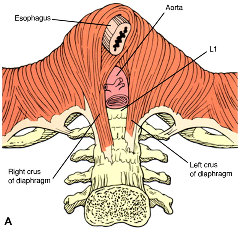
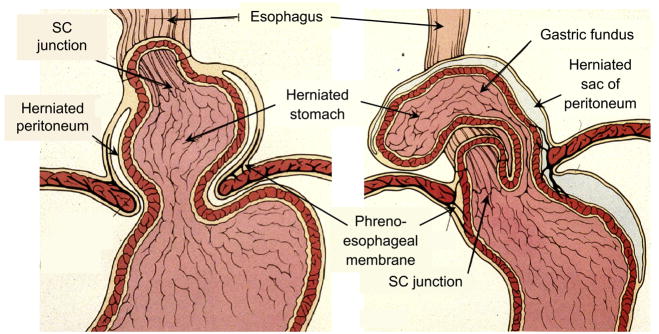
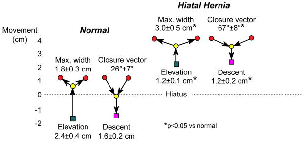
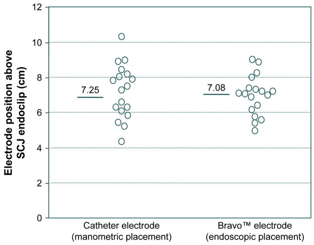
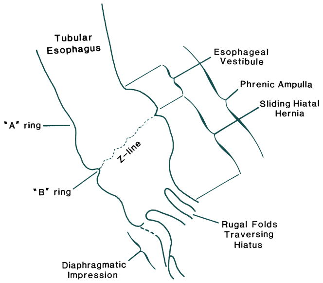
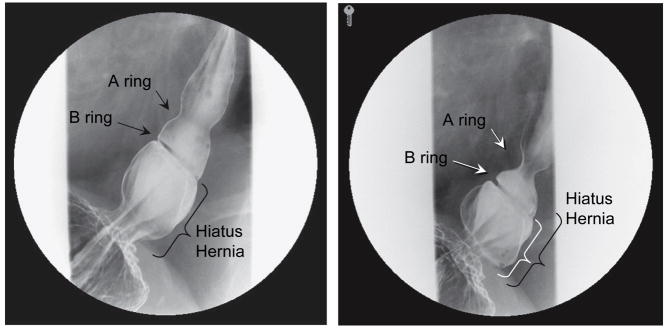
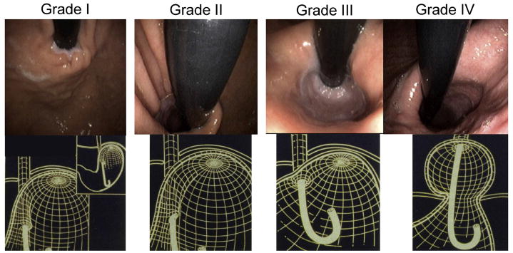
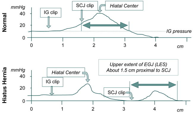
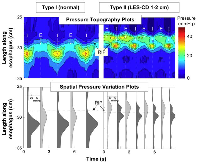
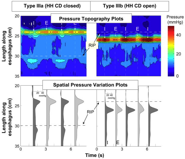
Hiatus hernia refers to conditions in which elements of the abdominal cavity, most commonly the stomach, herniate through the oesophageal hiatus into the mediastinum. With the most common type (type I or sliding hiatus hernia) this is associated with laxity of the phrenooesophageal membrane and the gastric cardia herniates. Sliding hiatus hernia is readily diagnosed by barium swallow radiography, endoscopy, or manometry when greater than 2 cm in axial span. However, the mobility of the oesophagogastric junction precludes the reliable detection of more subtle disruption by endoscopy or radiography. Detecting lesser degrees of axial separation between the lower oesophageal sphincter and crural diaphragm can only be reliably accomplished with high-resolution manometry, a technique that permits real time localization of these oesophagogastric junction components without swallow or distention related artefact.


Figures

Figure 1
Figure 1
The most common anatomy of the diaphragmatic hiatus in which the muscular elements of the crural diaphragm derive from the right diaphragmatic crus. The right crus arises from the anterior longitudinal ligament overlying the lumbar vertebrae. Once muscular elements emerge from the tendon, two flat muscular bands form that cross each other in scissor-like fashion, form the walls of the hiatus, and decussate with each other anterior to the esophagus. Modified from Jaffee BM, Surgery of the esophagus. In Orlando RC Ed. Atlas of Esophageal Diseases, Second Edition. pp 223–242.
Figure 2
Figure 2
Distinction between a sliding hiatal hernia (type I) and paraesophageal hernia (type II). With type I hernia the leading edge is the gastric cardia while with type two it is the gastric fundus. The SCJ maintains its native position in the paraesophageal hernia while it is displaced upward with the sliding hernia. Modified from Jaffee BM, Surgery of the esophagus. In Orlando RC Ed. Atlas of Esophageal Diseases, Second Edition. pp 223–242.
Figure 3
Figure 3
Axial mobility of the EGJ during swallow in normal controls and patients with type I hiatus hernia demonstrated by fluoroscopically tracking the movement of endoscopically place endoclips at the squamocolumnar junction. Clip movements are referenced to a point on the vertebral column. The green square represents the position of the SCJ prior to swallow, almost 2 cm distal to the center of the hiatus in the normal controls and 2 cm above it in the case of the hernia patients. With the initiation of swallow, the clip elevates to the point indicated by the yellow circle before luminal opening occurs. The red circles then indicate the maximal diameter of opening achieved, significantly wider in the case of the hernia patients, consistent with the reduced compliance demonstrable in these patients. During closure the angle of descent is steeper with the normals than with the hernia patients and the final position (magenta square) is essentially the same as prior to the swallow. Laxity and loss of elasticity of the phrenoesophageal membrane is suggested by two observations: 1) the attenuated axial movement of the SCJ in the hernia patients and 2) the flattening of the closure vector in the hernia patients suggesting diminished elastic recoil as the longitudinal muscle of the esophagus relaxes post-swallow. Modified from Kahrilas, PJ, Wu, S, Lin, S, et al. Attenuation of esophageal shortening during peristalsis with hiatus hernia. Gastroenterology 1995;109:1818.
Figure 4
Figure 4
Accuracy and variability in manometric and endoscopic pH electrode placement assessed by fluoroscopic imaging of final placement in reference to an endoclip placed at the SCJ. In all cases, the intended placement was 6 cm proximal to the SCJ. Data for the 18 subjects all of whom had both pH electrodes in place concurrently. Both intra-subject and inter-subject variability were greater with the manometric placement. Presumably, much of the inaccuracy in placement with both electrodes was attributable to shortening of the esophageal during the assessment of SCJ or LES position attributable to endoscopy or manometry. Modified from Pandolfino JE, Schreiner MA, Lee TJ, et al. Comparison of the Bravo Wireless and Digitrapper Catheter-based pH Monitoring Systems for Measuring Esophageal Acid Exposure. Am J Gastroenterol 2005;100:1466–1476.
Figure 5
Figure 5
Anatomical features of a sliding hiatus hernia viewed radiographically during swallowing. The A ring is a muscular ring visible during swallowing which demarcates the superior margin of the LES. Physiologically, the A ring corresponds to the highest pressure zone within the LES. The B ring at the squamocolumnar junction is present in only about 15% of individuals and allows for accurate division of the phrenic ampulla into the esophageal vestibule (A ring to B ring) and the sliding hiatus hernia (B ring to the subdiaphragmatic stomach). By convention the distinction between normal and hiatus hernia is a ≥ 2cm separation between the B ring and the hiatus. Rugal folds traversing the hiatus support the conviction that a portion of the stomach is supradiaphragmatic. From Kahrilas, PJ. Hiatus hernia causes reflux: fact or fiction? Gullet 1993;3(Suppl):21, with permission.
Figure 6
Figure 6
Radiographs taken sequentially during a swallow of a patient with a small axial hiatal hernia, a well developed A ring, and a well developed B ring evident. In such cases the criterion for defining hiatus hernia is that the measurement from the B ring to the diaphragmatic hiatus exceed 2 cm. This distance is indicated by the black bracket in the image on the left, obtained early in the swallow sequence, and the white bracket in the image on the right, obtained late in the swallow sequence. The black bracket from the left image is superimposed on the right making the point that size estimate of a sliding hiatus hernia will vary depending on when in the swallow sequence the measurement is made.
Figure 7
Figure 7
Endoscopic appearance and corresponding three-dimensional representation of the progressive anatomic disruption of the gastroesophageal junction as occurs with development of a Type I hiatus hernia. In the grade I configuration, a ridge of muscular tissue is closely approximated to the shaft of the retroflexed endoscope. With a grade II configuration the ridge of tissue is slightly less well defined and there has been slight orad displacement of the squamocolumnar junction along with widening of the angle of His. In the grade III appearance the ridge of tissue at the gastric entryway is barely present and there is often incomplete luminal closure around the endoscope. Note, however, that this is not a hiatal hernia because the SCJ is not displaced axially in the endoscopic photograph. With grade IV deformity, no muscular ridge is present at the gastric entry. The gastroesophageal area stays open all the time, and squamous epithelium of the distal esophagus can be seen from the retroflexed endoscopic view. A hiatus hernia is always present with grade IV deformity. Modified from Hill, LD, Kraemer, SJM, Aye, RW, et al. Laparoscopic Hill repair. Contemporary Surgery, 1994;44:1.
Figure 8
Figure 8
Examples of manometric pull-through tracings with single (top) and double (bottom) peak axial pressure profiles. In each case, an endoclip was placed at the intragastric (IG) aspect of the EGJ and at the SCJ before the subject underwent a pull through under fluoroscopy with a manometric catheter that also had radio-opaque markers at the pressure recording sites. This allowed for correlation between the clipped anatomic landmarks, the pressure profile, and the fluoroscopic landmarks localized on the pull through tracings. Note the 2 cm separation between the hiatal center and the intragastric clip in the normal; this corresponds to the “submerged segment” of the EGJ. Also note that the proximal aspect of the EGJ pressure profile extends 1.5 cm proximal to the EGJ, corresponding to position of the A ring often evident in barium swallow radiography. Data from: Kahrilas PJ, Lin S, Chen J, Manka M. The effect of hiatus hernia on gastro-oesophageal junction pressure. Gut 1999;44:483–489.
Figure 9
Figure 9
High resolution manometry examples of EGJ pressure morphology subtypes primarily distinguished by the extent of lower esophageal sphincter-crural diaphragm (LES-CD) separation. The upper plot in each panel is a pressure topography representation of the pressure changes spanning from the distal esophagus, across the EGJ, and into the proximal stomach during several respiratory cycles. The pressure scale is shown at the right. The lower plots illustrates a series of spatial pressure variation plots at the instants of peak inspiration (dark gray) and expiration (light gray) corresponding to the times marked I and E on the upper panels with pressure magnitudeon the x-axis and axial location along the y-axis. The location of the respiratory inversion point (RIP) is shown by the horizontal dashed line. Type I is characterized by complete overlap of the CD and the LES with a single pressure peak in the spatial pressure variation plots during both inspiration and expiration. The RIP lies at the proximal margin of the EGJ. Type II is characterized by minimal, but discernible, LES-CD separation making for a double peaked spatial pressure variation plot, but the nadir pressure between the peaks was still greater than gastric pressure. The RIP is within the EGJ at the proximal margin of the CD. Modified from: Pandolfino JE, Kim H, Ghosh SK, Clarke JO, Zhang Q, Kahrilas PJ. High resolution manometry of the EGJ: and analysis of crural diaphragm function in GERD. Am J Gastroenterol 2007;102:1056–1063.
Figure 10
Figure 10
Same layout as Figure 10. EGJ type IIIa was defined when LES-CD separation was >2 cm at inspiration. This is the high resolution manometry signature of hiatus hernia. Two subtypes were discernible, IIIa and IIIb, with the distinction being that the respiratory inversion point was proximal to the CD with IIIa and proximal to the LES in IIIb. The shift in respiratory inversion point is likely indicative of a grossly patulous hiatus, open throughout the respiratory cycle. Minimal EGJ pressure increase reflecting CD contraction is observed during inspiration with either type. Modified from: Pandolfino JE, Kim H, Ghosh SK, Clarke JO, Zhang Q, Kahrilas PJ. High resolution manometry of the EGJ: and analysis of crural diaphragm function in GERD. Am J Gastroenterol 2007;102:1056–1063.

References

    1. Marchand P. The anatomy of esophageal hiatus of the diaphragm and the pathogenesis of hiatus herniation. Thorac Surg. 1959;37:81–92. - PubMed
    1. Barrett NR. Discussion on hiatus hernia. Proc Roy Soc Med. 1932;122:736–796.
    1. Schatzki R. Die hernien des hiatus oesophageus. Deutsches Arch f klin med. 1932;173:85–103.
    1. Low A. A note on the crura of the diaphragm and the muscle of Treitsz. J Anatomy Lond. 1907;42:93–96. - PMC - PubMed
    1. Kahrilas PJ, Lin S, Chen J, Manka M. The effect of hiatus hernia on gastro-oesophageal junction pressure. Gut. 1999;44:483–489. - PMC - PubMed

Publication types