. 2008 Aug 19;47(33):8623-37.
doi: 10.1021/bi800873n. Epub 2008 Jul 26.

Purification and mutagenesis of LpxL, the lauroyltransferase of Escherichia coli lipid A biosynthesis

Affiliations

Purification and mutagenesis of LpxL, the lauroyltransferase of Escherichia coli lipid A biosynthesis

David A Six et al. Biochemistry. .

Abstract

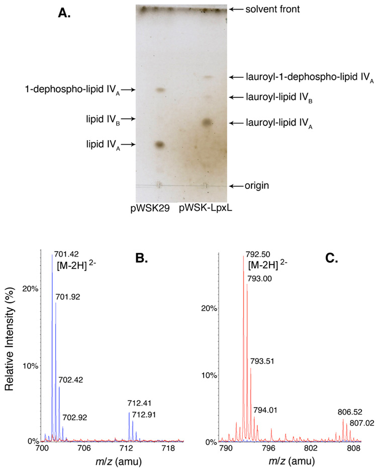
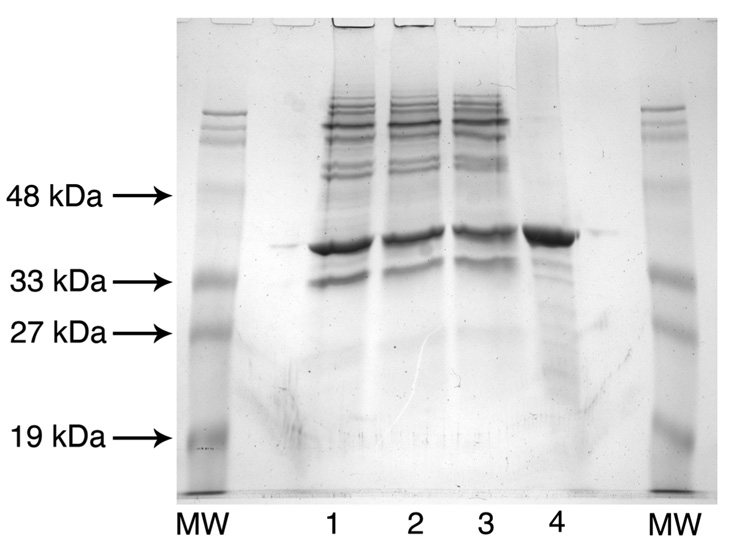
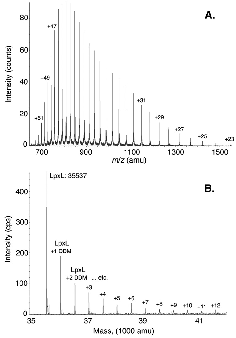
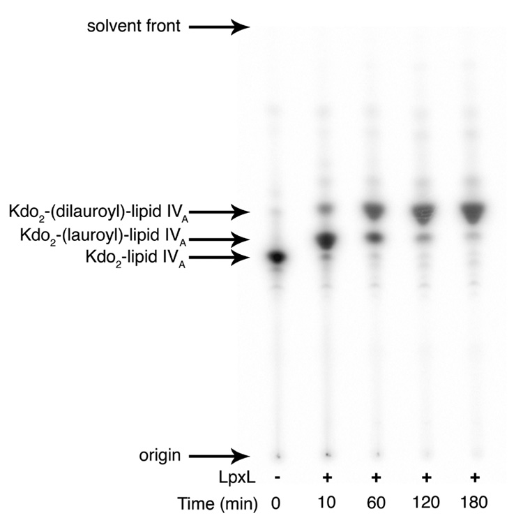
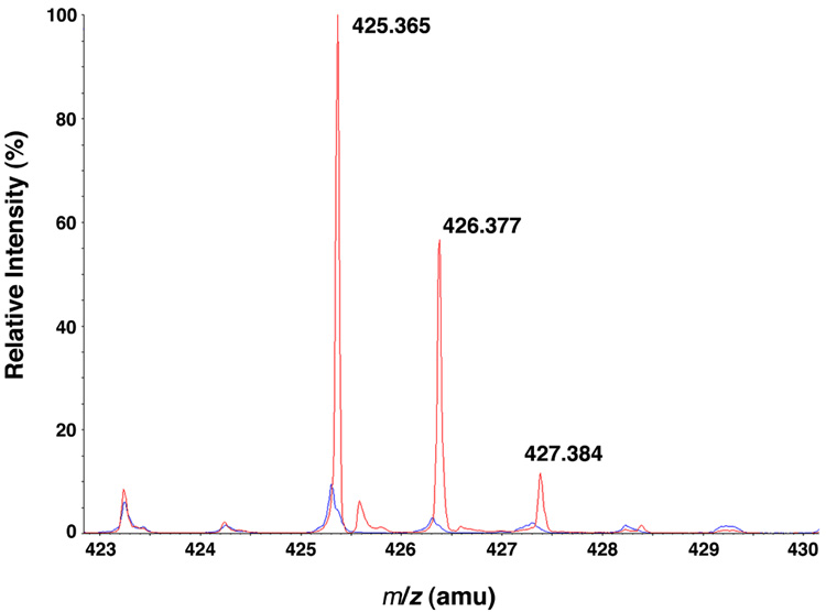
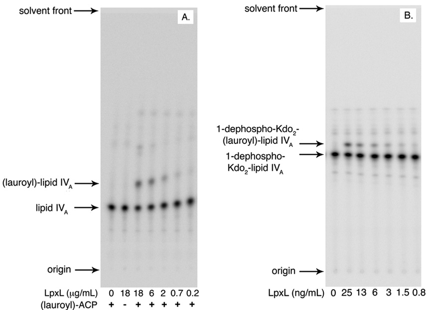
Escherichia coli lipid A is a hexaacylated disaccharide of glucosamine with secondary laurate and myristate chains on the distal unit. Hexaacylated lipid A is a potent agonist of human Toll-like receptor 4, whereas its tetra- and pentaacylated precursors are antagonists. The inner membrane enzyme LpxL transfers laurate from lauroyl-acyl carrier protein to the 2'- R-3-hydroxymyristate moiety of the tetraacylated lipid A precursor Kdo 2-lipid IV A. LpxL has now been overexpressed, solubilized with n-dodecyl beta- d-maltopyranoside (DDM), and purified to homogeneity. LpxL migration on a gel filtration column is consistent with a molecular mass of 80 kDa, suggestive of an LpxL monomer (36 kDa) embedded in a DDM micelle. Mass spectrometry showed that deformylated LpxL was the predominant species, noncovalently bound to as many as 12 DDM molecules. Purified LpxL catalyzed not only the formation in vitro of Kdo 2-(lauroyl)-lipid IV A but also a slow second acylation, generating Kdo 2-(dilauroyl)-lipid IV A. Consistent with the Kdo dependence of crude LpxL in membranes, Kdo 2-lipid IV A is preferred 6000-fold over lipid IV A by the pure enzyme. Sequence comparisons suggest that LpxL shares distant homology with the glycerol-3-phosphate acyltransferase (GPAT) family, including a putative catalytic dyad located in a conserved H(X) 4D/E motif. Mutation of H132 or E137 to alanine reduces specific activity by over 3 orders of magnitude. Like many GPATs, LpxL can also utilize acyl-CoA as an alternative acyl donor, albeit at a slower rate. Our results show that the acyltransferases that generate the secondary acyl chains of lipid A are members of the GPAT family and set the stage for structural studies.


Figures

Figure 1
Figure 1. An assay for LpxL in living cells of E. coli
A. The lipid A species released from the LPS of IPTG-induced mutant MKV15b (lacking lpxL, LpxM and LpxP), harboring either the vector control pWSK29 (left lane) or pWSK-LpxL (right lane), were subjected to TLC in the solvent chloroform:pyridine:88% formic acid:water (50:50:16:5, v/v) and visualized by charring. B. The lipid A species isolated from MKV15b cells harboring either the vector control pWSK29 or pWSK-LpxL were subjected to ESI/MS in the negative ion mode, and the spectra were normalized and overlayed. The major peak for the vector control sample (blue) was seen at m/z 701.42 and is interpreted as the [M-2H]2− ion of lipid IVA. The minor peak at m/z 712.41 is the corresponding sodium adduct [M-3H+Na]2−. The spectrum of lipid A species from MKV15b cells harboring pWSK-LpxL (red) shows no peaks in this range, consistent with the TLC analysis. C. The major peak for the lipid A species from MKV15b cells harboring the pWSK-LpxL (red) is seen at m/z 792.50, corresponding to the [M-2H]2− ion of (lauroyl)-lipid IVA. The minor peak at m/z 806.52 may correspond to (myristoyl)-lipid IVA, reflecting slightly relaxed LpxL substrate specificity. The spectrum of the vector control sample (blue) contains no peaks in this range.
Figure 2
Figure 2. Purification of LpxL to near homogeneity
This Coomassie-Blue-stained SDS polyacrylamide gel shows the degree of protein purity for each fraction assayed in Table II. Approximately 20 µg of protein was loaded in each lane. The molecular weight standards are present in the outside lanes, as indicated. Lane 1, membranes from induced MKV15b/pWSK-LpxL; Lane 2, 2% DDM-treated membranes before centrifugation; Lane 3, DDM-solubilized membranes (the high-speed supernatant of the DDM-treated membranes); and Lane 4, LpxL purified over a cellulose phosphate column, pooled, and concentrated. The molecular weight of LpxL was confirmed to be 35.5 kDa.
Figure 3
Figure 3. LpxL in vitro activity assay
Serial dilutions of pure LpxL (from 220 ng/mL to 0.1 ng/mL) were assayed in vitro with 6.25 µM Kdo2-[4′-32P]-lipid IVA (1,000 cpm/µL) and 12.5 µM lauroyl-ACP at 30 °C for 10 min in a final volume of 10 µL. The assay mixture also included 0.1 mg/mL BSA, 5 mM MgCl2, 50 mM NaCl, 0.1% Triton X-100, and 50 mM HEPES, pH 7.5. Portions of 4 µL were spotted on a silica TLC plate and developed with the solvent chloroform:pyridine:88% formic acid:water (30:70:16:5, v/v). The plates were dried and analyzed with a PhosphorImager system.
Figure 4
Figure 4. LC/ESI/MS of purified LpxL
Pure LpxL was subjected to LC/ESI/MS in the positive ion mode, as described in the Materials and Methods. A. LpxL elutes without DDM between 7 and 7.7 min. The peaks for the m/z range of 650 – 1550 amu are shown, corresponding to various charge states (H+) of LpxL ranging from +23 to +53. B. LpxL elutes with DDM between 7.72 and 8.10 min. The protein identification program parameters were limited to proteins in the molecular weight range of 33 – 43 kDa using the m/z range of 640 – 1020. Although protein ion peaks were identified above 1020, a cutoff of 1020 was selected because of an interfering protonated DDM dimer at m/z 1021.64. The mass reconstruction of the deconvoluted masses for LpxL and LpxL-DDM adducts is shown.
Figure 5
Figure 5. LpxL kinetics and detergent dependency
To determine the apparent Km and Vmax for each of its two substrates, LpxL was assayed at a fixed, saturating concentration of one substrate while the other was varied. The buffer included 0.1 mg/mL BSA, 5 mM MgCl2, 50 mM NaCl, and 50 mM HEPES, pH 7.5. Portions of 4 µL were spotted onto a silica TLC plate, which was developed in the solvent chloroform:pyridine:88% formic acid:water (30:70:16:5, v/v) and quantified with a PhosphorImager. A. LpxL at 4.7 ng/mL was assayed at 30 min with 75 µM Kdo2-[4′-32P]-lipid IVA (5,000 cpm/µL), 0.1% Triton X-100, and varying concentrations of lauroyl-ACP (3 to 180 µM) in a total volume of 10 µL for each lauroyl-ACP concentration. The specific activity was determined at each lauroyl-ACP concentration. The Michaelis-Menten equation was fit to the data to provide an apparent KM and Vmax. For lauroyl-ACP, the apparent KM is 7 ± 2 µM, and the apparent Vmax is 95 ± 5 µmol/min/mg. B. LpxL at a concentration of 0.18 ng/mL was assayed from 10 to 30 min with 90 µM lauroyl-ACP, 0.1% Triton X-100 and varying concentrations of Kdo2-[4′-32P]-lipid IVA (700 cpm/µL, 3 µM to 180 µM) in a total volume of 10 µL for each Kdo2-lipid IVA concentration. The specific activity was determined at each Kdo2-[4′-32P]-lipid IVA concentration. For Kdo2-lipid IVA the apparent KM is 15 ± 3 µM and the apparent Vmax is 221 ± 7 µmol/min/mg. C. The effects of varying Triton X-100 on LpxL activity were determined. LpxL was assayed for 10 min at 1.6 ng/mL at 15 µM for Kdo2-[4′-32P]-lipid IVA (1,000 cpm/µL) and 90 µM lauroyl-ACP in a total volume of 10 µL at each Triton X-100 concentration. The Triton X-100 concentration was varied from 0.0155 to 2%, and the fraction of substrate conversion to product was determined. The data points are connected for ease of visualization only.
Figure 6
Figure 6. Formation of Kdo2-[4′-32P]-(dilauroyl)-lipid IVA at high substrate and LpxL concentrations
A large excess of LpxL (17 µg/mL) was incubated with 100 µM Kdo2-[4′-32P]-lipid IVA (1,000 cpm/µL) and 300 µM lauroyl-ACP for 10 to 180 min in a total volume of 11 µL. The buffer included 0.1% Triton X-100, 0.1 mg/mL BSA, 5 mM MgCl2, 50 mM NaCl, and 50 mM HEPES, pH 7.5. At the given time points, a 2-µL portion was spotted onto a silica TLC plate, which was developed with chloroform:pyridine:88% formic acid:water (30:70:16:5, v/v) and analyzed with a PhosphorImager system.
Figure 7
Figure 7. Confirmation of a 3′-secondary lauroyl chain in LpxL-generated Kdo2-(dilauroyl)-lipid IVA
The Kdo2-(dilauroyl)-lipid IVA generated by LpxL was treated with membranes from either vector control cells or LpxR over-expressing cells. The lipids were extracted from the reaction mixture and fractionated on DEAE cellulose. The compounds that eluted with 30 mM ammonium acetate (42) were isolated and subjected to ESI/MS, and the spectra were normalized and overlayed. The LpxR-treated Kdo2-(dilauroyl)-lipid IVA released a lipid characterized by m/z 425.365 (red), which is interpreted as the [M-H] ion of the lauroyl ester of R-3-hydroxymyristic acid. The spectrum from the vector control-treated Kdo2-(dilauroyl)-lipid IVA (blue) does not contain this species.
Figure 8
Figure 8. Dependence of LpxL on the Kdo and 1-phosphate moieties
In vitro assays with these alternative substrates were performed at 30 °C in reaction mixtures containing 0.1 mg/mL BSA, 5 mM MgCl2, 50 mM NaCl, 0.1% Triton X-100, and 50 mM HEPES, pH 7.5. A. Serial dilutions of pure LpxL (from 18 µg/mL to 0.22 µg/mL) were assayed with 6.25 µM [4′-32P]-lipid IVA (1,000 cpm/µL) and 25 µM lauroyl-ACP at for 60 min in a total volume of 10 µL at each LpxL concentration. Portions of 4 µL were spotted on a silica TLC plate, which was developed with chloroform:pyridine:88% formic acid:water (50:50:16:10, v/v). B. Serial dilutions of pure LpxL (from 25 ng/mL to 0.8 ng/mL) were assayed with 1.25 µM 1-dephospho-Kdo2-[4′-32P]-lipid IVA (800 cpm/µL) and 12.5 µM lauroyl-ACP at for 30 min in a total volume of 10 µL at each LpxL concentration. Portions of 4 µL were spotted onto a silica TLC plate, which was developed with chloroform:pyridine:88% formic acid:water (30:70:16:5, v/v) and quantified with a PhosphorImager.
Figure 9
Figure 9. Lauroyl-CoA is both a slow substrate and an inhibitor of LpxL
Pure LpxL (40 ng/mL) was assayed with 6.25 µM Kdo2-[4′-32P]-lipid IVA (1,000 cpm/µL) and varying concentrations of lauroyl-CoA (from 1 µM to 500 µM) at 30 °C for 30 min in a total volume of 10 µL at each lauroyl-CoA concentration. The buffer included 0.1 mg/mL BSA, 0.5 mM LiCl, 5 mM MgCl2, 50 mM NaCl, 0.1% Triton X-100, and 50 mM HEPES, pH 7.5. Portions of 4 µL were spotted on a silica TLC plate, which was developed with chloroform:pyridine:88% formic acid:water (30:70:16:5, v/v) and analyzed with a PhosphorImager.
Scheme 1
Scheme 1. Incorporation of laurate into E. coli lipid A by LpxL
The 2′ secondary laurate chain incorporated by LpxL is shown in red in the context of the entire constitutive lipid A pathway. Other enzyme designations are shown in magenta. The glucosamine disaccharide of lipid A is highlighted in blue, with the numbering system indicated in red. LpxM does not function efficiently without the laurate chain in its substrate (23, 24).

References

    1. Raetz CRH, Whitfield C. Lipopolysaccharide endotoxins. Annu Rev Biochem. 2002;71:635–700. - PMC - PubMed
    1. Raetz CRH, Reynolds CM, Trent MS, Bishop RE. Lipid A modification systems in gram-negative bacteria. Annu Rev Biochem. 2007;76:295–329. - PMC - PubMed
    1. Miller SI, Ernst RK, Bader MW. LPS, TLR4 and infectious disease diversity. Nat Rev Microbiol. 2005;3:36–46. - PubMed
    1. Kim HM, Park BS, Kim JI, Kim SE, Lee J, Oh SC, Enkhbayar P, Matsushima N, Lee H, Yoo OJ, Lee JO. Crystal structure of the TLR4-MD-2 complex with bound endotoxin antagonist Eritoran. Cell. 2007;130:906–917. - PubMed
    1. Coats SR, Do CT, Karimi-Naser LM, Braham PH, Darveau RP. Antagonistic lipopolysaccharides block E. coli lipopolysaccharide function at human TLR4 via interaction with the human MD-2 lipopolysaccharide binding site. Cell Microbiol. 2007;9:1191–1202. - PubMed

Publication types

MeSH terms

LinkOut - more resources