. 2008 Oct;54(1):104-13.
doi: 10.1111/j.1574-695X.2008.00459.x. Epub 2008 Jul 24.

Chlamydiae and polymorphonuclear leukocytes: unlikely allies in the spread of chlamydial infection

Affiliations

Chlamydiae and polymorphonuclear leukocytes: unlikely allies in the spread of chlamydial infection

Roger G Rank et al. FEMS Immunol Med Microbiol. 2008 Oct.

Abstract

While much is known about the attachment of the chlamydiae to the host cell and intracellular events during the developmental cycle, little is known about the mechanism(s) by which elementary bodies exit the cell. In this report, we use the guinea-pig conjunctival model of Chlamydia caviae infection to present in vivo ultrastructural evidence supporting two mechanisms for release of chlamydiae from the mucosal epithelia. Four days after infection, histopathologic observation shows an intense infiltration of polymorphonuclear leukocytes (PMN) in the conjunctival epithelium. Using transmission electron microscopy, a gradient-directed PMN response to chlamydiae-infected epithelial cells was observed. As PMN infiltration intensifies, epithelial hemidesmosome/integrin/focal adhesion adherence with the basal lamina is disconnected and PMNs literally lift off and release infected superficial epithelia from the mucosa. Many of these infected cells appear to be healthy with intact microvilli, nuclei, and mitochondria. While lysis of some infected cells occurs with release of chlamydiae into the extracellular surface milieu, the majority of infected cells are pushed off the epithelium. We propose that PMNs play an active role in detaching infected cells from the epithelium and that these infected cells eventually die releasing organisms but, in the process, move to new tissue sites via fluid dynamics.


Figures
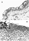
Figure 1
Figure 1
Hematoxylin and eosin-stained section of guinea pig conjunctiva showing the acute inflammatory response. (A) Control uninfected conjunctival epithelium showing goblet cells and a epithelium only a few cells in depth. (B) Inflamed epithelium showing heavy infiltration of the epithelium by PMNs. Occasional mononuclear cells are also seen in the submucosa. 40 ×. (Insert) High-powered view showing a chlamydia-infected cell (arrow) with a PMN (open arrow) directly underneath and in contact with the cell that is being dislodged from the epithelium.
Figure 2
Figure 2
Low power (10×) view of a section of infected epithelium stained with mouse anti-chlamydial LPS. The staining is restricted to the epithelium, demonstrating that the infection is restricted to superficial epithelial cells. Note two lymphoid aggregates (arrows) which are characteristic of the guinea pig conjunctiva. These are routinely found in infected animals as well.
Figure 3
Figure 3
PMN response to C. caviae infection of superficial epithelia cells of guinea pig conjunctiva. (A) Some of the prominent chlamydial inclusions are indicated by arrows and enlarged in panels B–D. (D) The infected epithelial cell cluster has apparently been released from the conjunctiva into the lumen. Serial sections of this cell confirmed that it is not attached to the epithelium. Stars denote the PMNs juxtaposed to some chlamydiae infected epithelial cells. Magnifications: A = × 2,500; B = × 17,900; C and D = × 12,500.
Figure 4
Figure 4
All stages of the chlamydial developmental cycle can be found in various infected conjunctival epithelial cells. (A) An EB and intermediate body; (B) an early inclusion containing a few RB; (C) a larger inclusion filled with RB and EB. The latter inclusion more clearly depicts the tendency of C. caviae to initially form multiple small inclusions, which may later fuse to form larger inclusions. Magnifications: A = × 25,100; B = × 17,900; C and D = × 12,500.
Figure 5
Figure 5
Results of the PMN response to chlamydiae-infected conjunctival epithelial cells. (A) PMNs apparently in the act of “pushing” the infected epithelial cells off the mucosal lining, causing a breech in the barrier with (B) subsequent release of intact, infected epithelial cells, damaged epithelial cells, chlamydiae (arrowheads) and PMNs. One can discern a progression in events (as denoted by the circled numbers) in which 1) PMNs accumulate under an intact infected epithelium; 2) the epithelial cell layer begins to lose its integrity; and 3) the epithelium has been breached, releasing PMNs onto the surface. Arrows denote examples of chlamydiae-infected superficial epithelial cells, which are more clearly evident in enlarged images in Figures 6A–C. Magnification: × 3,760.
Figure 6
Figure 6
Chlamydiae in inclusions in infected guinea pig conjunctival superficial epithelial cells abutted by PMNs. (A–C) Representative enlarged electron photomicrographs of portions of infected epithelial cells, designated by arrows in Figure 5, to more clearly reveal chlamydial morphology. (D) Enlarged electron photomicrograph of another example of intact infected epithelial cells seemingly being lifted off the conjunctival surface by underlying PMNs. Note that the desmosomes (black oval) are still intact at the epithelial cell lateral membranes. Tight junctions (black squares) are visible between the epithelial cells of the secondary layer; however, the inter-epithelial cell PMN has disrupted the left-most tight junction of the sub-epithelial cell. Furthermore, note that the detaching of the superficial epithelial cells has now exposed the microvilli of the underlying cells of the secondary layer. Magnifications: A–B = 7,530; C = 10,500; D = 12,500.
Figure 7
Figure 7
Chlamydiae can also be found in the extracellular environment and inside infiltrating PMNs. (A and inset) Released EBs/IBs (arrowheads) and RBs (arrows) were observed extracellularly as well as in apical PMNs. (B and C) A strong and specific immunogold labeling of EBs and RBs was detected when anti-chlamydial LPS polyclonal antibodies were exposed to the Lowicryl sections of infected epithelial cells. Magnifications: A = 7530; B = 25,100; C = 35,800; D = 17,900; E = 30,400.
Figure 8
Figure 8
Intact chlamydiae in vesicles within PMNs. (A,C) EB/IB (arrowheads) in inter-epithelial PMNs, enlarged, respectively, in (C and D). (E) Higher magnification of EBs (arrowheads) and an RB (arrow) in separate vesicles in a PMN. Magnifications: A and C = 7,000; B = 17,900; D = 25,000; and E = 30,400.

Similar articles

Cited by

References

    1. Abu El-Asrar AM, Geboes K, al Kharashi SA, Tabbara KF, Missotten L. Collagen content and types in trachomatous conjunctivitis. Eye. 1998;12(Pt 4):735–739. - PubMed
    1. Barnhart KT, Stolpen A, Pretorius ES, Malamud D. Distribution of a spermicide containing Nonoxynol-9 in the vaginal canal and the upper female reproductive tract. Hum.Reprod. 2001;16:1151–1154. - PubMed
    1. Barron AL, White HJ, Rank RG, Soloff BL. Target tissues associated with genital infection of female guinea pigs by the chlamydial agent of guinea pig inclusion conjunctivitis. J.Infect.Dis. 1979;139:60–68. - PubMed
    1. Barteneva N, Theodor I, Peterson EM, de la Maza LM. Role of neutrophils in controlling early stages of a Chlamydia trachomatis infection. Infection and Immunity. 1996;64:4830–4833. - PMC - PubMed
    1. Buchholz KR, Stephens RS. Activation of the host cell proinflammatory interleukin-8 response by Chlamydia trachomatis. Cell Microbiol. 2006;8:1768–1779. - PubMed

Publication types

MeSH terms