. 2009 Sep;13(9B):3753-63.
doi: 10.1111/j.1582-4934.2008.00430.x. Epub 2008 Jul 24.

Cyclooxygenase in normal human tissues--is COX-1 really a constitutive isoform, and COX-2 an inducible isoform?

Affiliations

Cyclooxygenase in normal human tissues--is COX-1 really a constitutive isoform, and COX-2 an inducible isoform?

Nina Zidar et al. J Cell Mol Med. 2009 Sep.

Abstract

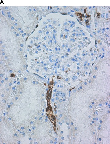
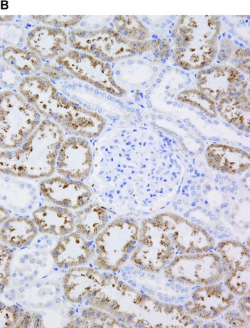
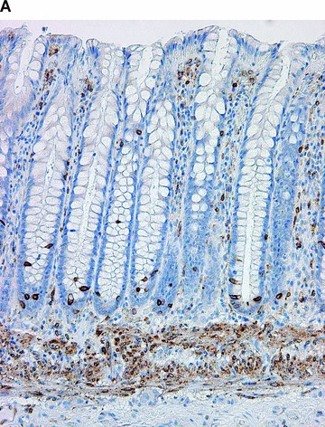
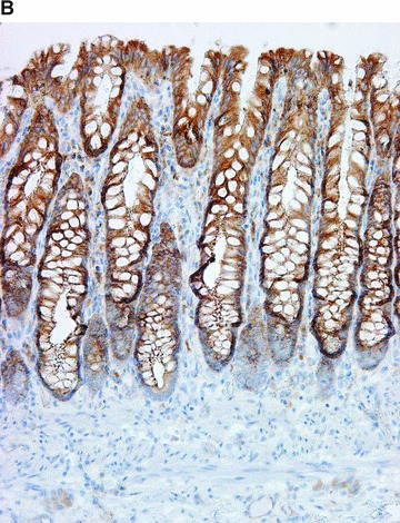
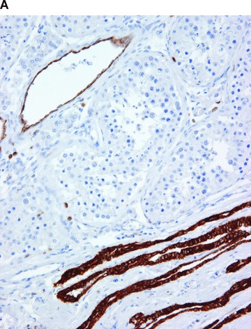
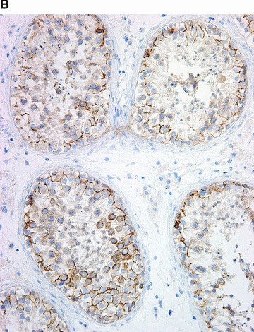
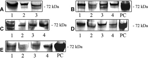
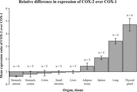
Cyclooxygenase (COX) is a key enzyme in prostanoid synthesis. It exists in two isoforms, COX-1 and COX-2. COX-1 is referred to as a 'constitutive isoform', and is considered to be expressed in most tissues under basal conditions. In contrast, COX-2 is referred to as an 'inducible isoform', which is believed to be undetectable in most normal tissues, but can be up-regulated during various conditions, many of them pathological. Even though the role of COX in homeostasis and disease in now well appreciated, controversial information is available concerning the distribution of COX isoforms in normal human tissues. There is mounting evidence that it is much more complex than generally believed. Our aim was therefore to analyse the expression and distribution of COX isoforms in normal human tissues, using immunohistochemistry, Western blotting and real-time RT-PCR. Autopsy samples from 20 healthy trauma victims and samples from 48 biopsy surgical specimens were included. COX-1 was found in blood vessels, interstitial cells, smooth muscle cells, platelets and mesothelial cells. In contrast, COX-2 was found predominantly in the parenchymal cells of many tissues, with few exceptions, for example the heart. Our results confirm the hypothesis that the distribution of COX isoforms in healthy tissues is much more complex than generally believed. This and previous studies indicate that both isoforms, not only COX-1, are present in many normal human tissues, and that both isoforms, not only COX-2, are up-regulated in various pathological conditions. We may have to revise the concept of 'constitutive' and 'inducible' COX isoforms.


Figures

Figure 1
Figure 1
COX in the brain. Positive immunohistochemical reaction for COX-1 in the glial cells (A), and for COX-2 in the neurons (B).
Figure 1
Figure 1
COX in the brain. Positive immunohistochemical reaction for COX-1 in the glial cells (A), and for COX-2 in the neurons (B).
Figure 2
Figure 2
COX in the lung. Positive immunohistochemical reaction for COX-1 in blood vessels and in smooth muscle cells in the bronchiolar wall (A). Positive immunohistochemical reaction for COX-2 in epithelial cells of the bronchiolar wall, and in inflammatory cells (B).
Figure 2
Figure 2
COX in the lung. Positive immunohistochemical reaction for COX-1 in blood vessels and in smooth muscle cells in the bronchiolar wall (A). Positive immunohistochemical reaction for COX-2 in epithelial cells of the bronchiolar wall, and in inflammatory cells (B).
Figure 3
Figure 3
COX in the kidney. Positive immunohistochemical reaction for COX-1 in the terminal portion of afferent arterioles at the glomerular entrance, in interstitial cells and in the parietal epithelial cells of the Bowman’s capsule (A). Positive immunohistochemical reaction for COX-2 in proximal tubules (B).
Figure 3
Figure 3
COX in the kidney. Positive immunohistochemical reaction for COX-1 in the terminal portion of afferent arterioles at the glomerular entrance, in interstitial cells and in the parietal epithelial cells of the Bowman’s capsule (A). Positive immunohistochemical reaction for COX-2 in proximal tubules (B).
Figure 4
Figure 4
COX in the large bowel. Positive immunohistochemical reaction for COX-1 in the muscularis mucosae, in crypt endocrine cells, and cells in the lamina propria (A). Positive immunohistochemical reaction for COX-2 in the surface and crypt epithelium, and rare cells in lamina propria (B).
Figure 4
Figure 4
COX in the large bowel. Positive immunohistochemical reaction for COX-1 in the muscularis mucosae, in crypt endocrine cells, and cells in the lamina propria (A). Positive immunohistochemical reaction for COX-2 in the surface and crypt epithelium, and rare cells in lamina propria (B).
Figure 5
Figure 5
COX in the testis. Positive immunohistochemical reaction for COX-1 in blood vessels, in epithelium of rete testis, and in rare interstitial cells (A). Positive immunohistochemical reaction for COX-2 in the seminiferous tubules (B).
Figure 5
Figure 5
COX in the testis. Positive immunohistochemical reaction for COX-1 in blood vessels, in epithelium of rete testis, and in rare interstitial cells (A). Positive immunohistochemical reaction for COX-2 in the seminiferous tubules (B).
Figure 6
Figure 6
Expression of COX-2 protein in normal human organs and tissues, as detected with Western blot analysis. (A); COX-2 in the endocrine glands: adrenal cortex (1), thyroid gland (2), hypophysis (3). (B); COX-2 in the liver and digestive tract: liver (1), mucosa of the colon (2), mucosa of the stomach – antrum (3), mucosa of the stomach – corpus (4). (C); COX-2 in the reproductive organs: testis (1), prostate (2), uterus (3) and periovulatory-phase ovary (4). (D); COX-2 in the kidney cortex (1), brain (2), lung (3) and spleen (4). (E); COX-2 in the cardiovascular system, where it was only occasionally detected in samples from the heart (1), aorta (2) and coronary artery (3). COX-2 in the subepicardial adipose tissue (4). PC; positive control (RAW 264.7 + LPS/PMA cell lysate).
Figure 7
Figure 7
Mean expression ratio of COX-2 over COX-1 (N± S.D.) in normal human organs and tissues as detected by real-time RT-PCR. In some organs (stomach, colon and small intestine), the expression of COX-1 mRNA was greater than the expression of COX-2 (N < 1: expression of COX-1 is 1/N-fold greater). In other organs (lung, thyroid gland, spleen, adipose tissue), the expression of COX-2 mRNA was greater than the expression of COX-1 (N > 1: expression of COX-2 is N-fold greater). In the liver, COX-1 and COX-2 mRNAs were equally expressed (N= 1). The differences in the expression level of the two target genes were statistically significant in the stomach (antrum), spleen, lung and thyroid gland (P < 0.05).

References

    1. Simmons DL, Botting RM, Hla T. Cyclooxygenase isozymes: the biology of prostaglandin synthesis and inhibition. Pharmacol Rev. 2004;56:387–437. - PubMed
    1. Anderson WF, Umar A, Viner JL, et al. The role of cyclooxygenase inhibitors in cancer prevention. Curr Pharm Des. 2002;8:1035–62. - PubMed
    1. Seibert K, Masferrer JL. Role of inducible cyclooxygenase (COX-2) in inflammation. Receptor. 1994;4:17–23. - PubMed
    1. Vane JR, Bakhle YS, Botting RM. Cyclooxygenases 1 and 2. Annu Rev Pharmacol Toxicol. 1998;38:97–120. - PubMed
    1. Cippolone F, Fazia ML. COX-2 and atherosclerosis. J Cardiovasc Pharmacol. 2006;47:S26–36. - PubMed