. 2008 Oct;213(4):391-424.
doi: 10.1111/j.1469-7580.2008.00953.x. Epub 2008 Jul 22.

From fish to modern humans--comparative anatomy, homologies and evolution of the head and neck musculature

Affiliations

From fish to modern humans--comparative anatomy, homologies and evolution of the head and neck musculature

R Diogo et al. J Anat. 2008 Oct.

Abstract

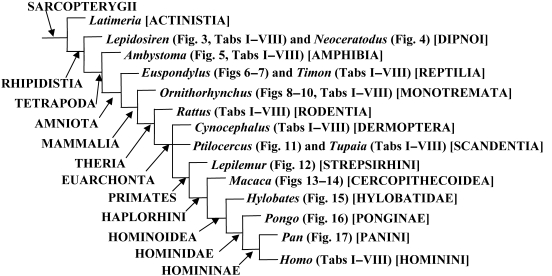
In a recent paper Diogo (2008) reported the results of the first part of an investigation of the comparative anatomy, homologies and evolution of the head and neck muscles of osteichthyans (bony fish + tetrapods). That report mainly focused on actinopterygian fish, but also compared these fish with certain non-mammalian sarcopterygians. The present paper focuses mainly on sarcopterygians, and particularly on how the head and neck muscles have evolved during the transitions from sarcopterygian fish and non-mammalian tetrapods to monotreme and therian mammals, including modern humans. The data obtained from our dissections of the head and neck muscles of representative members of sarcopterygian fish, amphibians, reptiles, monotremes and therian mammals, such as rodents, tree-shrews, colugos and primates, including modern humans, are compared with the information available in the literature. Our observations and comparisons indicate that the number of mandibular and true branchial muscles (sensu this work) present in modern humans is smaller than that found in mammals such as tree-shrews, rats and monotremes, as well as in reptiles such as lizards. Regarding the pharyngeal musculature, there is an increase in the number of muscles at the time of the evolutionary transition leading to therian mammals, but there was no significant increase during the transition leading to the emergence of higher primates and modern humans. The number of hypobranchial muscles is relatively constant within the therian mammals we examined, although in this case modern humans have more muscles than other mammals. The number of laryngeal and facial muscles in modern humans is greater than that found in most other therian taxa. Interestingly, modern humans possess peculiar laryngeal and facial muscles that are not present in the majority of the other mammalian taxa; this seems to corroborate the crucial role played by vocal communication and by facial expressions in primate and especially in human evolution. It is hoped that by compiling, in one paper, data about the head and neck muscles of a wide range of sarcopterygians, the present work could be useful to comparative anatomists, evolutionary biologists and functional morphologists and to researchers working in other fields such as developmental biology, genetics and/or evolutionary developmental biology.


Figures

Fig. 1
Fig. 1
Phylogenetic framework for the discussion provided in the present paper and the comparison between the head and neck muscles of the genera listed in Tables 1–4 and shown in Figs 3–17, based on Shoshani et al. (1996), Kardong (2002), Sargis (2002a,, , Dawkins (2004), Kemp (2005), Marivaux et al. (2006), Diogo (2007), Janeka et al. (2007), and Silcox et al. (2007). N.B. When we use the term reptiles we refer to the group including taxa such as turtles, tuataras, lizards, snakes, crocodiles, and Aves, which, despite some controversy, continues to be considered a monophyletic taxon by most taxonomists and in most general textbooks (e.g. Kardong, 2002; Dawkins, 2004; Diogo, 2007); the Primates, Dermoptera (including colugos or ‘flying lemurs’) and Scandentia (including tree-shrews) are placed in an unresolved trichotomy, because the relationships between these three groups remains mainly unresolved (some authors continuing to group colugos with tree-shrews, others group tree-shrews with primates, and yet others group colugos with primates: e.g. Sargis, 2002a,b, 2004; Dawkins, 2004; Marivaux et al. 2006; Janeka et al. 2007; Silcox et al. 2007).
Fig. 3
Fig. 3
Lepidosiren paradoxa (Dipnoi): A) lateral view of the cephalic musculature; B) ventral view of the cephalic musculature (modified from Bemis & Lauder, 1986 and Diogo, 2008; the nomenclature of the structures illustrated follows that used in the present work; anterior is to the right). ADM, adductor mandibulae complex; CM, coracomandibularis; DM, depressor mandibulae; HYP, hypaxialis; INTE, interhyoideus; INTM, intermandibularis; part, prearticular; RE-AO, retractor anguli oris; SH, sternohyoideus.
Fig. 17
Fig. 17
Pan troglodytes(Mammalia, Primates): A) lateral view of the laryngeal musculature; B) same view, but the thyrohyoideus, sternothyroideus, constrictor pharyngis inferior and cricothyroideus were removed and the lateral portions of the thyroid cartilage and hyoid bone were partially cut [modified from Starck & Schneider (1960) and Saban (1968); the nomenclature of the structures illustrated basically follows that used in the present work; anterior is to the top, dorsal is to the right: see text]. ARY, arytenoideus; CRAL, cricoarytenoideus lateralis; CRAP, cricoarytenoideus posterior; cric, cricoid cartilage; CRTO, CRTR, pars obliqua and pars recta of cricothyroideus; epigl, epiglottis; IPC, constrictor pharyngis inferior; STT, sternothyroideus; THAR, thyroarytenoideus; THH, thyrohyoideus; thyr, thyroid cartilage.
Fig. 2
Fig. 2
Schematic presentation of embryonic origin of cranial muscles in gnathostomes based on Edgeworth's (1902, 1911, 1923, 1926a,b,c, 1928, 1935) works: premyogenic cells originate from the paraxial mesoderm (hatched areas) and several somites (areas with vertical bars); large arrows indicate a contribution of cells in segments of the mesoderm to muscle formation of different cranial arches. For more details, see text (modified from Miyake et al., 1992; the nomenclature of the structures illustrated follows that of these authors).
Fig. 5
Fig. 5
Ambystoma ordinarium (Amphibia): ventral view of the cephalic musculature; on the right side the most ventral muscles were removed (modified from Diogo, 2008; the nomenclature of the structures illustrated follows that used in the present work; anterior is to the top). BH, branchiohyoideus; ch, ceratohyal; DM-A, DM-P, depressor mandibulae anterior and posterior; GG, genioglossus; GH, geniohyoideus; INTE-A, INTE-P, anterior and posterior bundles of interhyoideus; INTM-A, INTM-P, intermandibularis anterior and posterior; l-ch-mand, ligament between ceratohyal and mandible; mnd, mandible; OH, omohyoideus; SAR1, subarcualis rectus 1; SH, sternohyoideus; uh, urohyal.
Fig. 9
Fig. 9
Ornithorhynchus anatinus(Mammalia, Monotremata): A) ventral view of the head and neck musculature, muscles such as the geniohyoideus and sternohyoideus are not shown; B) same view, but the digastricus anterior, superficial part of the mylohyoideus, sternomastoideus, and cleidomastoideus were removed and the anterior portion of the sternohyoideus and of the superficial part of the omohyoideus were partially cut (modified from Edgeworth, 1935 and Saban, 1971; the nomenclature of the structures illustrated basically follows that used in the present work; anterior is to the top). CMA, cleidomastoideus; DETR, detrahens mandibulae; DIA, digastricus anterior; GEH, geniohyoideus; MA, masseter; mnd, mandible; MYHPR, MYHSU, pars profunda and pars superficialis of mylohyoideus; OM, omohyoideus; OMPR, OMSU, pars profunda and pars superficialis of omohyoideus; SMA, sternomastoideus; STEH, sternohyoideus; STT, sternothyroideus; STYH, styloideus.
Fig. 16
Fig. 16
Pongo pygmaeus(Mammalia, Primates): ventral view of the head musculature; on the right side the superficial portion of the masseter was removed (modified from Edgeworth, 1935 and Saban, 1968; the nomenclature of the structures illustrated basically follows that used in the present work; anterior is to the top). DIP, digastricus posterior; mnd, mandible; MA, masseter; MPT, pterygoideus medialis; MYH, mylohyoideus; STG, styloglossus; STH, stylohyoideus.
Fig. 4
Fig. 4
Neoceratodus forsteri (Dipnoi): mesial view of adductor mandibulae and mandible; the mandibular tooth-plates are not illustrated (modified from Diogo, 2008; the nomenclature of the structures illustrated follows that used in the present work; anterior is to the right, dorsal is to the top). A2, A2-PVM, A3′, adductor mandibulae A2, A2-PVM and A3′; ang, angular; ar-pq, articulatory facet for palatoquadrate; part, prearticular; sppsp, splenio-postsplenial bone.
Fig. 6
Fig. 6
Euspondylus acutirostris (Reptilia): lateral view of the cephalic musculature; the adductor mandibulae A2-PVM is not shown (modified (2002 and Diogo, 2008; the nomenclature of the structures illustrated follows that used in the present work; anterior is to the right). A2, adductor mandibulae A2; CERV, cervicomandibularis; DM, depressor mandibulae; LEV-AO-M, levator anguli oris mandibularis; PSEU, pseudotemporalis.
Fig. 7
Fig. 7
Euspondylus acutirostris(Reptilia): ventral view of the deep ventral cephalic musculature; muscles such as the intermandibularis, interhyoideus, geniohyoideus, genioglossus and hyoglossus are not shown (modified from Montero et al. 2002 and Diogo, 2008; the nomenclature of the structures illustrated follows that used in the present work; anterior is to the top). cb1, ceratobranchial 1; CEH, ceratohyoideus; ehy, epihyal; hc, hyoid cornu; HYOB, hyobranchilais; mnd, mandible; OH, omohyoideus; pte, pterygoid; PTM, pterygomandibularis; SH, sternohyoideus.
Fig. 13
Fig. 13
Macaca mullata(Mammalia, Primates): lateral view of the masseter and temporalis (modified from Schumacher, 1961 and Saban, 1968; the nomenclature of the structures illustrated basically follows that used in the present work; anterior is to the right). MA, masseter; TEMP, temporalis; zyar, zygomatic arch.
Fig. 12
Fig. 12
Lepilemur sp. (Mammalia, Primates): lateral view of the facial musculature; muscles such as the buccinatorius, orbicularis oris and mentalis are not shown (modified from Jouffroy & Saban, 1971; the nomenclature of the structures illustrated basically follows that used in the present work; anterior is to the right). AUOR, auriculo-orbitalis; AUS, auricularis superior; FRO, frontalis; MAAU, mandibulo-auricularis; NASL, nasolabialis; OCC, occipitalis; OROC, orbicularis oculi; PLAC, PLAM, platysma cervicale and platysma myoides; SCOP, sphincter colli profundus; ZYMA, ZYMI, zygomaticus major and zygomaticus minor.
Fig. 8
Fig. 8
Ornithorhynchus anatinus(Mammalia, Monotremata): lateral view of the deep facial musculature; muscles such as the interhyoideus profundus, buccinatorius, orbicularis oris and mentalis are not shown (modified from Lightoller, 1942 and Saban, 1971; the nomenclature of the structures illustrated basically follows that used in the present work; anterior is to the right). CETR, cervicalis transversus; OROC, orbicularis oculi; PLAC, PLAM, platysma cervicale and platysma myoides; SCOS, sphincter colli superficialis.
Fig. 14
Fig. 14
Macaca cyclopis(Mammalia, Primates): anterior view of the facial musculature; muscles such as the buccinatorius, platysma, frontalis, and occipitalis are not shown; on the left side the depressor supercilii was removed and the orbicularis oculi, zygomaticus minor, zygomaticus major, levator labii superioris, levator labii superioris alaeque nasi were partially cut (modified from Shibata, 1959 and Jouffroy & Saban, 1971; the nomenclature of the structures illustrated basically follows that used in the present work). COS, corrugator supercilii; DES, depressor supercilii; DLI, depressor labii inferioris; DSN, depressor septi nasi; LAO, levator anguli oris facialis; LELS, levator labii superioris; LELSA, levator labii superioris alaeque nasi; MENT, mentalis; NAS, nasalis; OROC, orbicularis oculi; OROR, orbicularis oris; PRO, procerus; ZYMA, ZYMI, zygomaticus major and zygomaticus minor.
Fig. 11
Fig. 11
Ptilocercus lowii(Mammalia, Scandentia): ventral view of the musculature of the hyoid region of the right side of the body; muscles such as the geniohyoideus, sternothyroideus and thyrohyoideus are not shown (modified from Le Gros Clark, 1926 and Saban, 1968; the nomenclature of the structures illustrated basically follows that used in the present work; anterior is to the right). aub, auditory bulla; CEH, ceratohyoideus; CNVII, X, XII, cranial nerves VII, X and XII; cric, cricoid cartilage; CRT, cricothyroideus; DIP, digastricus posterior; GEG, genioglossus; HYG, hyoglossus; OM, omohyoideus; SMA, sternomastoideus; STEH, sternohyoideus; STG, styloglossus; STH, stylohyoideus; STP, stylopharyngeus; thyr, thyroid cartilage.
Fig. 15
Fig. 15
Hylobates hoolock(Mammalia, Primates): lateral view of the pharyngeal musculature (modified from Kanagasuntheram, 1952–1954 and Saban, 1968; the nomenclature of the structures illustrated basically follows that used in the present work; anterior is to the top, dorsal is to the left: see text). CRT, cricothyroideus; GEG, genioglossus; HYG, hyoglossus; IPC, constrictor pharyngis inferiori; LVP, levator veli palatini; MGP, mylo-glossopharyngeus; MPC, constrictor pharyngis medius; PAP, palatopharyngeus; PTEP, pterygopharyngeus; STG, styloglossus; STP, stylopharyngeus.
Fig. 10
Fig. 10
Ornithorhynchus anatinus(Mammalia, Monotremata): dorsal view of the laryngeal musculature; the cricoarytenoideus posterior is not shown (modified from Göppert, 1937 and Saban, 1971; the nomenclature of the structures illustrated basically follows that used in the present work; anterior is to the top). ARY, arytenoideus; aryt, arytenoid cartilage; copth, copula thyroidea; corthI, cornua thyroidea I; cric, cricoid cartilage; epigl, epiglottis; mupr, muscular process; paryt, proarytenoid cartilage; TCAR, thyrocricoarytenoideus.

Similar articles

Cited by

References

    1. Abdala V, Moro S. A cladistic analysis of ten lizard families (Reptilia: Squamata) based on cranial musculature. Russ J Herpetol. 2003;10:53–78.
    1. Adamicka P, Ahnelt H. Two jaw articulations in Latimeria chalumnae(Actinistia, Coelacanthidae) Zool Jb Anat. 1992;122:107–112.
    1. Adams SB, Wheeler JFG, Edgeworth FH. On the innervation of the platysma and the mandibulo-auricularis. J Anat. 1929;63:242–252. - PMC - PubMed
    1. Alexander RMcN. Jaw mechanisms of the coelacanth Latimeria. Copeia. 1973;1973:156–158.
    1. Allis EP. The cranial muscles and cranial nerves of Amia calva. J Morphol. 1897;12:487–737.

Publication types

LinkOut - more resources