Comparative Study
. 2008 Jul 25:6:34.
doi: 10.1186/1741-7007-6-34.

Platypus globin genes and flanking loci suggest a new insertional model for beta-globin evolution in birds and mammals

Affiliations
Comparative Study

Platypus globin genes and flanking loci suggest a new insertional model for beta-globin evolution in birds and mammals

Vidushi S Patel et al. BMC Biol. .

Abstract

Background: Vertebrate alpha (alpha)- and beta (beta)-globin gene families exemplify the way in which genomes evolve to produce functional complexity. From tandem duplication of a single globin locus, the alpha- and beta-globin clusters expanded, and then were separated onto different chromosomes. The previous finding of a fossil beta-globin gene (omega) in the marsupial alpha-cluster, however, suggested that duplication of the alpha-beta cluster onto two chromosomes, followed by lineage-specific gene loss and duplication, produced paralogous alpha- and beta-globin clusters in birds and mammals. Here we analyse genomic data from an egg-laying monotreme mammal, the platypus (Ornithorhynchus anatinus), to explore haemoglobin evolution at the stem of the mammalian radiation.

Results: The platypus alpha-globin cluster (chromosome 21) contains embryonic and adult alpha- globin genes, a beta-like omega-globin gene, and the GBY globin gene with homology to cytoglobin, arranged as 5'-zeta-zeta'-alphaD-alpha3-alpha2-alpha1-omega-GBY-3'. The platypus beta-globin cluster (chromosome 2) contains single embryonic and adult globin genes arranged as 5'-epsilon-beta-3'. Surprisingly, all of these globin genes were expressed in some adult tissues. Comparison of flanking sequences revealed that all jawed vertebrate alpha-globin clusters are flanked by MPG-C16orf35 and LUC7L, whereas all bird and mammal beta-globin clusters are embedded in olfactory genes. Thus, the mammalian alpha- and beta-globin clusters are orthologous to the bird alpha- and beta-globin clusters respectively.

Conclusion: We propose that alpha- and beta-globin clusters evolved from an ancient MPG-C16orf35-alpha-beta-GBY-LUC7L arrangement 410 million years ago. A copy of the original beta (represented by omega in marsupials and monotremes) was inserted into an array of olfactory genes before the amniote radiation (>315 million years ago), then duplicated and diverged to form orthologous clusters of beta-globin genes with different expression profiles in different lineages.


Figures

Figure 1
Figure 1
Current proposed model for the evolution of α- and β-globin clusters from paralogous clusters in different lineages. The unlinked α- and β-globin clusters in birds and mammals evolved from an ancient in trans duplication of the ancestral linked α-β cluster, followed by differential gene silencing (marked with X). This resulted in bird β-like globin genes (β2) orthologous to the marsupial ω-globin gene (β2 beside the α-globin cluster) but paralogous to mammalian β-like globin genes (β1). Adapted from Wheeler et al. [36].
Figure 2
Figure 2
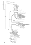
Evolutionary relationships among vertebrate α-like globin genes using a 50% majority rule consensus phylogram from an analysis using Bayesian Inference. The tree was constructed using mixed models of evolution for each codon position (see methods) and estimated base frequencies in an unlinked analysis using MrBayes (v. 3.1.2). Numbers adjacent to branches refer to % posterior probabilities. GenBank accession numbers for sequences are: Virginian Opossum (Didelphis virginiana) ζ1, ζ2, α1, α2, θ [AC139599.2, AC148752.1]; Stripe-faced Dunnart (Sminthopsis macroura) αD, α2, θ [AC146781]; Brazilian Opossum (Monodelphis domestica) α [TI# 453585430]; Tammar wallaby (Macropus eugenii) θ [AY459590], α [AY459589]; ζ [AY789121], ζ' [AY789122]; Horse (Equus caballus) θ (ψ α) [Y00284], α1 [M17902], ζ [X07051]; pig (Sus scrofa) αD [AC145444]; cat (Felis catus) αD [AC130194]; cow (Bos taurus) αD [AC150547]; Goat (Capra hircus) α [J00043]; Human (Homo sapiens) α1 [V00491], θ [X06482], ζ [NM_005332]; muD chain [AY698022]; Mouse (Mus muscularis) α1 [NM_008218], ζ [X62302]; Rabbit (Oryctolagus cuniculus)α [X04751]; Eastern Quoll (Dasyurus viverrinus) α [M14567]; Chicken (Gallus gallus) αA, π, αD [AF098919]; Duck (Cairina moschata) αD [X01831]; Pigeon (Columba livia) αD [AB001981]; Turtle (Geochelone nigra) αD [SEG# AB1165195]; Zebrafish (Danio rerio) α1 [NM_131257]; Salamander (Hynobius retardatus) larval α [AB034756]; Salamander (Pleurodeles waltlii) α [M13365]; Frog (Xenopus laevis) α I [X14259], larval (tadpole) α T5 [X02798]; Yellowtail (Seriola quinqueradiata) αA [AB034639]; Salmon (Salmo salar) α [X97289]; Southern Puffer (Sphoeroides nephelus) α2 [AY016023]; Platypus (Ornithorhynchus anatinus) ζ, ζ', αD, α3, α2, α1 [AC203513].
Figure 3
Figure 3
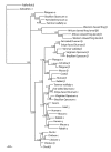
Evolutionary relationships among vertebrate β-like globin genes using a 50% majority rule consensus phylogram from an analysis using Bayesian inference. The tree was constructed using mixed models of evolution for each codon position (see methods) and estimated base frequencies in an unlinked analysis using MrBayes (v. 3.1.2). Numbers adjacent to branches refer to % posterior probabilities. GenBank accession numbers for sequences are: Fat-tailed Dunnart (Sminthopsis crassicaudata) β [Z69592], ε [Z48632], ω [AY014770]; Stripe-faced Dunnart (S. macroura) β, ε [AC148754]; Virginian Opossum (Didelphis virginiana) β [J03643], ε [J03642]; Brazilian Opossum (Monodelphis domestica) β [XM_001365299], ε [XM_001364448], ω [XM_001364828]; Tammar Wallaby (Macropus eugenii) β [AY450928], ε [AY450927], ω [AY014769]; African clawed frog (Xenopus laevis) larval β I [NM_001086273], larval βII [NM_001088028]; Western clawed frog (X. tropicalis) β [NM_203528], larval ε1 [NM_001016495]; Chicken (Gallus gallus) β (βA) [NM_205489], ε [NM_001004390], γ (βA) [NM_001031489]; Duck (Cairina moschata) β [J00926], ε [X15740]; Human (Homo sapiens) β [NM_000518], γ [BC130459], ε [NM_005330]; Mouse (Mus musculus) β (β1) [NM_008220], γ (β h0) [NW_001030869], ε (εy) [M26897]; Goat (Capra hirus)β A) [DQ350619], ε (εI) [X01912], γ [M15388]; Rabbit (Oryctolagus cuniculus) β, γ, ε [M18818]; Echidna (Tachyglossus aculeatus) β [L23800]; Pufferfish (Fugu rubripes) β [AY170464]; Zebrafish (Danio rerio) ε1 [NM_001103130]; Platypus β, ε [AC192436], ω [AC203513].
Figure 4
Figure 4
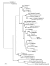
Evolutionary relationships among vertebrate β-like globin genes analysed by maximum parsimony (MP) trees of length 926 (one of eight trees). Third position in codons were excluded in the MP analyses, which were conducted using a heuristic search in PAUP* v.4.0b10 [65]. The tree is rooted using pufferfish β-globin. Numbers adjacent to branches represent % bootstrap values (>50%) from MP heuristic analyses of 1000 pseudoreplicates. Accession numbers for sequences are given in the caption of Figure 3.
Figure 5
Figure 5
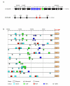
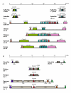
Gene structure of the platypus α- and β-globin clusters and flanking loci, and comparisons of their promoter regions with other mammals. (A) The platypus α-globin cluster contains six α-like globin genes (red), a β-like (ω) globin gene (blue) and a distantly related globin gene, GBY (green), which are flanked by IL9RP3-POLR3K-C16orf33-C16orf8-MPG-C16orf35 on the 5' end and LUC7L-ITFG3-RGS11-ARHGDIG-PDIA2-AXIN1 on the 3' end (black). The platypus β-globin cluster contains only two genes, ε and β (blue), which are flanked on both sides by ORG genes (black). (B) Relative positions of the putative transcription factor binding sites in the 200 bp promoter region located upstream of the predicted platypus, marsupial (Didelphis virginiana ζ and ψζ', and Sminthopsis macroura αD, ψα3, α2, α1, ω, ε and β) and human α- and β-like globin genes. For the platypus GBY no data was available from other species, including Xenopus tropicalis, for comparisons.
Figure 6
Figure 6
Expression of all predicted α- and β-like globin genes including GBY in an adult platypus. For each of the platypus predicted genes, expression was investigated by reverse transcriptase polymerase chain reaction in adult liver, kidney, spleen, testis, brain and lung. Primers for each gene were designed between two exons so that it would result in a product distinguishable from genomic contamination of cDNA. The negative control (last lane) contained no cDNA. All genes were expressed in one or more tissues, indicating that they are transcriptionally active and might be functional.
Figure 7
Figure 7
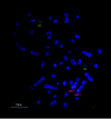
Chromosomal location of the platypus α- and β-globin clusters. Two-colour fluorescence in situ hybridisation showing the location of the α-globin cluster on chromosome 21 (green) and the β-globin cluster on chromosome 2q5.1 (red). The chromosomes are counterstained with DAPI (blue).
Figure 8
Figure 8
Loci flanking vertebrate α-globin (A) and β-globin (B) clusters. The relative locations of flanking loci (A) MPG, C16orf35, LUC7L and GBY and (B) RRM1, CCKBR, ILK and ORG genes were searched for beside the α-β globin cluster in zebrafish (Danio rerio) and frog (Xenopus tropicalis), and beside the separate α-globin and β-globin clusters in chicken (Gallus gallus), opossum (Monodelphis domestica) and human (Homo sapiens) from Ensembl [57]. The pufferfish (Fugu rubripes) flanking loci shown here were adapted from Gillemans et al. [8]. For the platypus, the α-globin flanking loci were characterised in this study, and ORG genes surrounding the platypus β-globin cluster were discovered: however, the BAC clone (484F22) was too small to cover the region containing the loci RRM1, CCKBR and ILK. In X. tropicalis LUC7L was found on another scaffold (466 from Ensembl) but sequence analyses by Fuchs et al. [2] suggested that LUC7L resides 3' to the frog α-β-GBY cluster. The flanking loci as well as the α- and β-globin clusters are differentiated by colour.
Figure 9
Figure 9
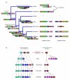
Proposed model for the evolution of the α- and β-globin clusters in vertebrate lineages. (A) A region containing MPG-C16orf35-α-β-GBY-LUC7L represented the ancient α-β globin cluster of jawed vertebrates (>450 MYA), which is seen in the amphibian lineage. This region further duplicated and underwent some gene silencing in teleost fish. In an amniote ancestor of reptiles, birds and mammals (>315 MYA), a copy of an ancestral β-globin gene from this region was inserted into a different chromosome within a region replete with multiple copies of ORG genes. The original amniote β-globin gene survives as the ω-globin gene (β1) in the α-globin cluster of marsupials and monotremes, whereas the transposed β-globin gene (β2) duplicated several times to form different clusters in the different lineages. (B) Tandem duplications of the ancestral amniote α-globin gene produced a three-gene (π-αDA) cluster in the avian lineage. In the mammalian lineage, further duplications gave rise to a six-gene (ζ-ζ'-αD321) cluster with ongoing gene conversion events homogenising the embryonic and adult genes. In monotremes, the ancestral ω (β1) and GBY are retained. After the divergence of monotreme and therian mammals, there was an additional duplication of α2 to form θ, giving rise to the seven-gene cluster (ζ-ζ'-αD321-θ) in marsupials and eutherians. Marsupials also retain the ancestral ω but may have lost GBY gene; eutherians retain no identifiable remnant of either gene. Furthermore, the ancestral transposed β2-globin gene duplicated independently in birds and mammals. Before the mammalian radiation, we propose that the ancestral β2 gene duplicated to form a two-gene β-globin cluster (ε-β) as seen in monotremes and marsupials, except that ongoing gene conversion events homogenised platypus ε to group with monotreme β genes. After the divergence of marsupial and eutherian mammals, there were further tandem duplications of these two genes to produce complex β-globin cluster (ε-γ-η-δ-β) in eutherians.

Similar articles

Cited by

References

    1. Roesner A, Fuchs C, Hankeln T, Burmester T. A globin gene of ancient evolutionary origin in lower vertebrates: evidence for two distinct globin families in animals. Mol Biol Evol. 2005;22:12–20. doi: 10.1093/molbev/msh258. - DOI - PubMed
    1. Fuchs C, Burmester T, Hankeln T. The amphibian globin gene repertoire as revealed by the Xenopus genome. Cytogenet Genome Res. 2006;112:296–306. doi: 10.1159/000089884. - DOI - PubMed
    1. Dickerson RE, Geis I. Hemoglobin: Structure, Function, Evolution and Pathology. Menlo Park, CA: Benjamin Cummings; 1983. pp. 65–116.
    1. Hentschel CC, Kay RM, Williams JG. Analysis of Xenopus laevis globins during development and erythroid cell maturation and the construction of recombinant plasmids containing sequences derived from adult globin mRNA. Dev Biol. 1979;72:350–363. doi: 10.1016/0012-1606(79)90124-6. - DOI - PubMed
    1. Jeffreys AJ, Wilson V, Wood D, Simons JP, Kay RM, Williams JG. Linkage of adult alpha- and beta-globin genes in X. laevis and gene duplication by tetraploidization. Cell. 1980;21:555–564. doi: 10.1016/0092-8674(80)90493-6. - DOI - PubMed

Publication types

LinkOut - more resources